New publications
Pelvic X-rays in women, men and children
Last reviewed: 06.07.2025

All iLive content is medically reviewed or fact checked to ensure as much factual accuracy as possible.
We have strict sourcing guidelines and only link to reputable media sites, academic research institutions and, whenever possible, medically peer reviewed studies. Note that the numbers in parentheses ([1], [2], etc.) are clickable links to these studies.
If you feel that any of our content is inaccurate, out-of-date, or otherwise questionable, please select it and press Ctrl + Enter.
Radiography as a method of visualizing various structures of the body, including pelvic X-rays, is crucial for identifying and correctly diagnosing injury, disease or pathology, as well as monitoring the results of their treatment. [ 1 ]
Indications for the procedure
The need for an X-ray examination of this anatomical area arises in patients admitted to emergency departments with severe injuries (as a result of a bruise, fall, traffic accident, etc.) and suspected fracture/crack of the pelvic bones (ilium, pelvic, pubic, sciatic); fracture of the pelvic ring, acetabulum or sacrum; dislocation or fracture of the hip joint. [ 2 ]
X-rays also help to identify the causes:
- pain in the pelvic bones and joints, including those caused by bone damage and degenerative diseases, as well as inflammation of the sacroiliac joint - sacroiliitis;
- pelvic pain, including chronic pelvic pain;
- pain in the groin area;
- pain in the pubic area.
To determine the condition of bone tissue and identify their destructive changes, an X-ray of the pelvis is performed for bedsores, which in bedridden patients form over the sacrum, coccyx, ischium or greater trochanter. Bedsores are especially dangerous due to infectious damage to the tissue of the underlying bones with the development of osteomyelitis. But X-ray diagnostics of osteomyelitis, as a rule, reveals bone damage in the late stages of the pathological process. Therefore, in this case, MRI is more informative, which allows you to obtain detailed images of both bones and surrounding soft tissues.
A pelvic x-ray (pelvis minor) – the pelvic cavity below the edges of the pelvic bones that contains the bladder and rectum – can detect later stages of cysts, tumors and pelvic bone infections.
Preparation
How to properly prepare for an X-ray of the pelvic bones? For several days, you should avoid eating foods containing fiber, which provoke increased formation of intestinal gases. The last meal should be no less than 10-12 hours before the procedure, in the morning you should do a cleansing enema.
In case of constipation, bowel cleansing before a pelvic X-ray is performed using laxatives, which are taken for three to four days before visiting the X-ray room.
Technique pelvic x-rays
Immediately before the X-ray, the patient's upper abdominal cavity is protected from X-rays with lead plates.
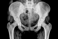
X-rays of the pelvic bones and hip joints can be taken in three projections: anteroposterior (AP), posteroanterior (PA), and lateral (side). The image from the front shows the pelvic bones from the upper part of the iliac crest to the proximal part of the femur shaft, the pubic and ischial bones, the hip joint, the obturator foramina, and the greater trochanters of the proximal femur in profile.
The lateral view shows the sacrum, coccyx, lumbosacral junction, superimposed femurs and upper thigh.
What does a pelvic x-ray show?
By visualizing the iliac crest, sacrum, sacroiliac joints, greater pelvic ring, pubic and ischium bones, proximal femur, pelvic X-ray can show: anatomical abnormalities of the pelvis or hip joint; pelvic fractures; fracture, dislocation or arthritis of the hip joint; tumors of the pelvic bones (osteosarcomas).
X-ray signs of a pelvic fracture see more in the publication X-ray signs of bone and joint damage
Also read – X-ray signs of bone and joint diseases
In addition, a woman's pelvic X-ray visualizes the uterus and its cervix, fallopian tubes and ovaries, i.e. the organs of the female reproductive system, located in the pelvis. And a man's pelvic X-ray shows the prostate gland and seminal glands (vesicles). But for diagnostic visualization of the listed pelvic organs, as well as the bladder, ultrasound is used, in particular: abdominal ultrasound of the pelvic organs and uterus, transvaginal ultrasound of the uterus, Doppler sonography of the fallopian tubes, ultrasound and transrectal ultrasound (TRUS) of the prostate, ultrasound of the bladder. And for examining the rectum, there are endoscopic methods - rectoscopy and colonoscopy.
Complications after the procedure
Although any exposure to ionizing radiation carries some risk to the body, diagnostic radiography is generally safe because it uses strictly dosed radiation. Read more in the article - Radiography