Medical expert of the article

New publications
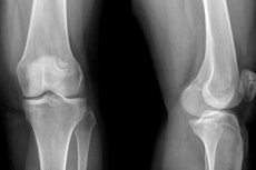
X-ray of the knee joint in two projections
Last reviewed: 04.07.2025

All iLive content is medically reviewed or fact checked to ensure as much factual accuracy as possible.
We have strict sourcing guidelines and only link to reputable media sites, academic research institutions and, whenever possible, medically peer reviewed studies. Note that the numbers in parentheses ([1], [2], etc.) are clickable links to these studies.
If you feel that any of our content is inaccurate, out-of-date, or otherwise questionable, please select it and press Ctrl + Enter.
Knee pain, impaired joint mobility in this area and traumatic injuries are quite common reasons for visiting a doctor. Even an experienced doctor cannot easily determine by eye what the unpleasant symptoms are associated with. But a diagnosis is not an essay on a free topic, and when making it, a surgeon, traumatologist or orthopedist must rely on accurate information that can be obtained by conducting additional diagnostic measures. One of such mandatory and inexpensive studies is an X-ray of the knee joint.
 [
[ Indications for the procedure
X-ray examination is a procedure that allows the doctor to see deep structures invisible to the human eye, which cannot illuminate soft tissues to examine denser formations. Unlike a fluorogram, which must be taken regularly once a year, a doctor prescribes an X-ray of the knee joint only if there is a suspicion of certain pathologies affecting the bones, cartilage tissue, and ligamentous apparatus of the knee. This usually happens when contacting a doctor about pain and limited movement of the leg or when admitted to the emergency room due to an injury.
What disorders and pathologies may require X-ray confirmation:
- Violations of the integrity of the joint bones. Hard tissues - bones - are best seen on X-ray images, it is clear that such a study makes it possible to accurately diagnose any damage to them: fractures, cracks and dents formed as a result of a strong blow. The value of an X-ray study in this case is also that the doctor receives information about the exact location of the damage, the location of bone fragments, the size of cracks and bone depressions.
- Dislocation/subluxation of the joint. The nature of the displacement can be judged by the incorrect positioning of the bones relative to each other. In a joint, the convexity of one bone should coincide with the depression in the other. Any misalignment may indicate a displacement of the bones as a result of a blow or careless movement.
- Damage to the ligamentous apparatus (ruptures, stretches). Their presence is judged by the distance between the bones, because the ligaments themselves do not fully reflect X-rays, so they are poorly visible.
- Traumatic injuries to the kneecap (patella) and meniscus (inner and outer cartilage). Also detected by bone displacement or cracks in it
- Congenital pathologies of bones and joints (osteodystrophy and osteopathy).
X-ray examination allows for an accurate diagnosis in the following cases:
- arthritis and arthrosis (changes in the shape of the joint and the size of the joint space are observed),
- osteoporosis and osteomyelitis (bone density changes in different areas, unusual layers may appear),
- synovitis (due to the accumulation of fluid in the joint and the increase in the thickness of the synovial capsule, the joint space increases),
- osteochondropathy of Koenig and Osgood-Schlatter (foci of bone necrosis with smooth, uneven edges are detected).
X-ray of the knee joint can also reveal pathologies that the patient did not even suspect. For example, tumor processes affecting the bones and soft tissues of the joint, the presence of cysts and unusual bone growths (osteophytes), the presence of a foreign body.
A visit to the doctor with complaints of pain in the area and changes in the shape of the knee (regardless of whether the person had an injury), impaired mobility of the knee joint, swelling and redness of soft tissues indicating an inflammatory process are already compelling reasons for prescribing an X-ray examination.
What does an X-ray of the knee joint show?
Preparation
X-ray of the knee joint is considered a procedure that does not require any preparation. A person can go for the examination immediately after consulting a doctor. X-ray of various parts of the lower limb does not require restrictions in nutrition and medication. And even if it is carried out with contrast. The fact is that the contrast is not injected into a vein, but directly into the joint capsule. The only thing that may be needed is an allergy test to identify the body's sensitivity to the contrast.
Before the procedure, it is advisable to expose the area being examined, as clothing may contain details that distort the radiographic image. If the patient previously had a bandage applied to the knee area, there is no need to remove it, but the devices that fix the leg in the desired position after the injury will have to be removed, if possible.
Since the lower part of the body is exposed to radiation, a special lead apron is first applied to the area of the reproductive organs, which does not allow X-rays to pass through. However, this is more relevant for children, whose body size is smaller than that of adults, which means that X-rays can also capture a small part of the child's body.
Technique knee X-rays
An X-ray of the knee joint of an infant (and it may be necessary due to birth injuries and congenital pathologies) is performed with the utmost caution. At the same time, the entire body of the baby is covered with special protective devices. This is due not only to the fact that radiation is more dangerous for a baby than for an adult. The growth of an infant is still very small, so the entire body of the child, and not just the limb being examined, can fall into the field created by the X-ray emitter.
There are no special nuances to radiography. The main requirement is to be in a static position as indicated by the doctor. Any movement will cause distortions in the images, making diagnostics difficult. Often in such cases, repeated radiography is required, and this is an additional dose of X-ray radiation.
It is most difficult for a child to remain still, so the X-ray table is equipped with special fixators. If pain is the cause of concern, the patient may be given an anesthetic injection to conduct a quality examination.
For accurate diagnosis of the above-described pathologies, usually not one, but at least two images in different projections are required. Direct projection (the image is taken when the person is lying on his back) is the most indicative when there is a suspicion of fractures of the bones entering the joint. In a standing position, several images can be taken: in the lateral, tangential and transcondylar projection. The latter, if necessary, can be done in a lying position on the side.
With tangential projection, pathologies of the patella and inflammatory-degenerative changes in the joints are better detected. Transcondylar projection is prescribed to detect ligament sprains, necrotic processes in bone tissue, and suspected osteoarthrosis. But with lateral projection, it is possible to diagnose fluid accumulation in the joints.
In some cases, doctors limit themselves to one projection, but in case of a controversial diagnosis, it is still more relevant to examine images taken from different angles. Most often, doctors prescribe an X-ray of the knee joint in two projections.
The performance of various structures of the knee joint can be assessed by taking additional pictures of the leg bent at different angles. In this case, radiography can be performed both at rest and with a load.
Contraindications to the procedure
X-ray of the knee joint is a procedure associated with the process of irradiation of the patient's limb with harmful ionizing radiation. However, if you cover the body with protective clothing, the consequences after the procedure will be minimal.
It is believed that X-ray irradiation has a negative impact on human health. However, this does not include symptoms typical for the early period after receiving a dose of radiation: reddening of the skin (radiation burn), epidermal detachment, the appearance of erosions, increased fatigue, etc. However, various sources incessantly talk about late complications after the procedure, such as an increased risk of cancer, mutational changes, decreased sexual function, etc.
In fact, such consequences are possible if you undergo X-rays daily for a long period without protective equipment. But according to reviews from doctors and patients, they have not encountered anything like this (at least, it was not possible to establish a clear relationship between the symptoms that appeared later and the diagnostic measures).
The radiation dose in modern X-ray machines during examination of the knee joint is approximately equal to the radiation dose that we receive in a day and a half of life in natural conditions. At the same time, it is tens of times less than that which surrounds us in airports and airplanes. Therefore, even repeated images are not capable of causing much harm to the body, even taking into account the radiation received while watching TV, working on a computer, etc.
However, the procedure does have some contraindications. It is not recommended for pregnant women and nursing mothers, because radiation can negatively affect the development of the fetus in the womb and penetrate into breast milk, and with it into the body of the newborn. If there is no other alternative, the woman's entire body, except for the knee, should be protected from the penetration of X-rays.
X-rays also have a negative effect on sperm quality, so you should abstain from sexual intercourse for some time after the procedure, the purpose of which is to conceive a child. However, X-ray results in obese people may be unreliable due to the high density of fatty tissue, which makes the images unclear.
It is not advisable to prescribe an X-ray examination to people diagnosed with schizophrenia, as well as to patients who are in very serious condition with signs of blood loss.
If diagnostics are performed on a child, it is better to choose safer methods if possible. The most popular diagnostic methods are ultrasound, computer tomography and magnetic resonance imaging. The safest of all is still considered to be MRI, where magnetic field energy is used instead of X-rays.
All these methods can be prescribed in combination with an X-ray or instead of it. When choosing what is better: ultrasound, CT or MRI, you need to understand that the difference between the studies is not only in safety for the body.
If the patient is faced with a choice of what to do, MRI or X-ray of the knee joint, it is necessary to understand that in case of pathologies of hard tissues, X-ray examination is preferable, i.e. a regular X-ray of the joint or computed tomography, which is also based on the penetrating ability of X-rays. At the same time, CT is considered more informative in case of injuries and neoplasms in the knee area.
But MRI easily helps diagnose diseases associated with soft tissue structures: muscles, cartilage, ligaments, i.e. tissues with a high water content, which reacts to the magnetic field.
True, the cost of computed tomography and magnetic resonance imaging is significantly higher than a simple X-ray, which is considered quite sufficient for diagnosing knee joint pathologies.
When choosing an ultrasound or X-ray of the knee joint, you should remember that the latter, although less safe, is more informative for diagnosing bone pathologies. If we are talking about the ligamentous apparatus, pathologies of the synovial bags and cartilage, it is better to give preference to an ultrasound examination, the cost of which is still lower than that of the popular MRI.
Normal performance
It should be said that the information from the X-ray image is intended primarily for specialists and has no value for a person far from the issues of anatomy. In the best case, the patient will be able to independently diagnose a bone fracture. In fact, the decoding of the information from the image should be entrusted to a doctor.
X-rays have good penetrating ability, but tissues of different density retain radiation to different degrees. Dense tissues absorb more rays, so they are more clearly visible on an X-ray image. Tendon and cartilage tissues are considered the most penetrable. The latter are not visible on the image at all, but their condition and characteristics can be judged by the size of the joint space (the larger the gap between the bones, the thicker the cartilage tissue) and the change in the relative position of the endplates.
When examining the X-ray results of a healthy knee joint closely, the distal parts of the femur and tibia, the kneecap (patella bone) and a small area of the fibula are clearly visible. All bones have approximately the same color, which indicates equal tissue density, as well as smooth surfaces without any defects (clearly defined areas of darkening or lightening, incomprehensible layers, changes in bone shape). Dark areas may indicate fractures and cracks, and too light areas correspond to tumors, cysts, fluid accumulation.
The heads of the bones correspond to the depressions, the joint space has normal dimensions, while its width on both sides of the joint should be the same, and the shape is symmetrical. The norm in the X-ray of the knee joint does not provide for any inclusions in the cavity of the joint space (growths, incomprehensible particles).
The meniscus of the knee joint is not visible on an X-ray, because it is cartilaginous tissue. The condition of such tissue can only be judged by the width of the joint space, as well as by the size and shape of a small wedge-shaped shadow, the expanded part of which should be directed downwards. If a meniscus injury is suspected, the purpose of an X-ray examination is to exclude or confirm a bone fracture in this area.
Well, with fractures, dislocations (when there is a displacement of bones relative to each other), tumors, everything seems to be clear, but how to determine inflammatory-dystrophic changes in tissues on an X-ray. Let's consider what signs on the image help the doctor make an accurate diagnosis:
X-ray signs of knee joint arthrosis. In this case, the assessment of the width of the joint space, which is examined in direct and lateral projections, comes to the fore. With arthrosis, the joint space narrows along the entire perimeter or in a separate area. The disease is characterized by thinning of the periosteum, on which X-rays can reveal tuberosities and sharpenings characteristic of growing osteophytes. At later stages of the disease, marginal compactions of the tissues of the articular bones may be noted.
X-ray signs of arthritis of the knee joint. Unlike arthrosis, which is considered an age-related degenerative disease, arthritis can remind of itself at a young age. In addition to the dystrophy of joint tissues characteristic of arthrosis, this disease is characterized by a local inflammatory process, reinforced by other disorders in the body.
The initial stages of the disease cannot be seen on an X-ray, but later on such signs as osteoporosis of the bones (a decrease in their density, as a result of which the color of hard tissues will be darker than usual), narrowing of the joint space characteristic of arthrosis and arthritis, the appearance of bone growths on the distal parts of the bones will appear. The surface of the joints gradually becomes flatter, bone and cartilage tissues change their structure and characteristics, gradually impairing the mobility of the joint (in this case, the joint space may be practically invisible).
X-ray signs of bursitis of the knee joint. The pathology consists of the development of an inflammatory process in the synovial bags of the knee. X-ray will show the deep location of these structures and areas of calcification, characteristic of the inflammatory process. In this case, narrowing of the joint space is not observed.
In fact, radiography is an additional method of diagnosing this disease. Its purpose is to exclude inflammatory-degenerative pathologies of the joints (arthrosis and arthritis), as well as traumatic injuries that cause pain in the knee.
X-ray signs of synovitis of the knee joint. Synovitis is a less known pathology than others, characterized by the accumulation of fluid in the joint cavity. In this case, an unusual darkening will be observed in the area of the synovial bag. In the chronic course of the pathology, thinning of the cartilaginous tissue and complete loss of cartilage are observed, holes are formed on the bone in the joint area, through which exudate flows into the cavity of the soft tissues. In this case, the formation of osteophytes is not observed.
X-ray signs of Baker's cyst of the knee joint. On the X-ray, the cyst looks like a rounded neoplasm of a light shade localized in the popliteal fossa, which is clearly visible in the lateral projection. In this case, doctors pay special attention to the clearly defined boundaries of the defect, characteristic of cystic formations.
Tumors on the image do not have clear boundaries and a specific shape. X-rays allow us to detect such neoplasms, but cannot say anything about their nature.
Care after the procedure
X-ray examination, despite all the dangers of X-ray radiation, is a painless procedure. The doctor spends just over 3-5 minutes on it, and a person can receive the results almost immediately.
If radiography is performed digitally, a person can receive the answer immediately on a disk or flash drive, and the information received can be viewed on a computer monitor. The clarity and contrast of a digital image is usually higher than that of an X-ray film. Even soft tissue structures can be viewed on it at the appropriate resolution.
It takes time (about 10 minutes) for the film image to develop in analog radiography, after which the patient receives the image. If an additional description of the image is required, you will have to wait some more time.
Some sources recommend drinking more fresh cow's milk after an X-ray procedure, as it helps remove radiation from the body. It must be said that there is no particular need for this, but given all the beneficial properties of a natural product, why not follow the advice that helps saturate the body with useful substances.
X-ray of the knee joint is a diagnostic procedure that allows doctors to make an accurate diagnosis of many diseases of the musculoskeletal system. This method is time-tested, affordable and relatively safe, given the low dose of radiation received during one X-ray session. The information obtained by the X-ray machine is considered sufficient for diagnosing most traumatic and inflammatory-degenerative diseases of the knee. And only in the case of inflammatory and oncological processes may additional diagnostic methods be required.