Medical expert of the article

New publications
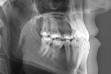
Adult and child jaw x-rays
Last reviewed: 03.07.2025

All iLive content is medically reviewed or fact checked to ensure as much factual accuracy as possible.
We have strict sourcing guidelines and only link to reputable media sites, academic research institutions and, whenever possible, medically peer reviewed studies. Note that the numbers in parentheses ([1], [2], etc.) are clickable links to these studies.
If you feel that any of our content is inaccurate, out-of-date, or otherwise questionable, please select it and press Ctrl + Enter.
Radiography in medicine is a method of studying the anatomical structures of the body to obtain their projection using X-rays on paper or film, which does not require penetration inside. It is difficult to imagine modern diagnostics without it. X-ray of the jaw allows dentists, maxillofacial, plastic surgeons to make a correct diagnosis and monitor treatment.
Digital radiography was introduced in the mid-1980s [ 1 ] and with continued growth in popularity it now competes with traditional screen film radiography (SFR) in all radiographic applications. [ 2 ]
Indications for the procedure
Examination of the patient allows the doctor to make assumptions about the diagnosis, but only an X-ray will give an accurate picture and choice of treatment algorithm.
Indications for its implementation are:
- in dentistry - problems with teeth, bone tissue, gums (caries, inflammation, abscess, periodontal disease, cysts and tumor processes, osteomyelitis, etc.), the result of filling, installation of implants, jaw prostheses, braces;
- in maxillofacial and plastic surgery - determining the extent and nature of damage in various injuries, improving appearance.
X-ray of an adult's jaw
What is revealed by an X-ray of the jaw in an adult? In addition to the listed dental diagnoses, these may be various defects (fractures, cracks, fragments), sclerotic processes, areas of dead tissue, bone growths and other pathological changes.
The need for X-rays during pregnancy (due to a lack of calcium during this period, teeth suffer greatly) often causes concern among expectant mothers who are concerned about the health of their child.
Modern equipment allows you to do an X-ray examination quite safely. The radiovisiograph, which the X-ray machine is equipped with, acts specifically on a specific tooth, has low radiation, and displays a clear picture on the monitor. And yet, in the first trimester of pregnancy, it is best to refrain from this procedure.
X-ray of a child's jaw
Despite the small doses of radiation, small children are very sensitive to X-rays, their internal organs are located closer, so it is better to protect them and not perform the procedure until 3-4 years. Orthopantogram or panoramic dental X-ray is recommended to be done no earlier than 5 years.
When does it become necessary to take a picture of babies? In addition to cases of injury, it is used to monitor the growth of teeth, the eruption of permanent teeth, align them, prevent the development of bone tissue diseases, and assess the condition of the oral cavity.
Technique jaw X-rays
For a complete picture of the jaw condition, several projections are required. Thus, an X-ray of the lower jaw is performed in the direct and lateral directions. The first provides general information, the second - the condition of the desired side. The technique of the procedure does not cause difficulties.
Direct projection is obtained in a horizontal position. The person is placed on his stomach face down, with the tip of the nose and forehead resting on the cassette, and the X-ray sensor located on the side of the occipital protuberance.
Lateral is done lying on the side, the cassette is placed under the cheek at a slight angle. Sometimes an axial (transverse) section is also required. In this case, the patient lies on his stomach, the head is pulled forward as much as possible, and the cassette is held by the neck and lower jaw.
X-ray of the upper jaw consists of two images: with the mouth closed and open. The body is on the stomach, the chin and the tip of the nose touch the cassette, the sensor is perpendicular to it.
3d x-ray of jaw
Since digital radiography has found its application in dentistry, many new applications for medical imaging have been proposed, including dental image registration, lesion detection, bone healing analysis, osteoporosis diagnosis, and dental forensics.[ 3 ]
Computer tomography or 3D X-ray allows you to make a high-quality volumetric image of the jaw in any projection, to create a 3D model of the jaw. Without carrying out traumatic procedures, this method makes it possible to obtain a virtual tissue section and look into any of their layers.
This procedure cannot be avoided when planning bone grafting, implantation or augmentation of the maxillary sinus floor.
Panoramic x-ray of the jaw
Panoramic radiography is currently the most widely used extraoral technique in modern dentistry due to its low cost, simplicity, information content and reduced impact on the patient. Since this radiographic method gives the dentist a general view of the alveolar process, condyles, sinuses and teeth, it plays a major role in the diagnosis of caries, jaw fractures, systemic bone diseases, unobstructed teeth and intraosseous lesions.
This type of examination is called an orthopantomogram and is a circular x-ray of the jaw. The information obtained in this way is called a dental passport. For a dentist, it reveals data on the presence and location of carious cavities, evaluates bone tissue for suitability for implantation, detects anomalies, inflammation, and poor-quality fillings.
The image can be viewed on a screen, enlarged, stored on a storage medium, or taken as a photograph. Successful panoramic radiography requires careful patient positioning and proper technique. [ 4 ] Proper technique requires the patient to be in an upright position with the neck extended, shoulders down, back straight, and feet together. [ 5 ]
X-ray of the jaw with baby teeth
In pediatric dentistry, X-rays are an integral part of diagnostics. Although baby teeth are temporary, they need to be treated in order for healthy permanent teeth to form.
On the eve of therapy, an X-ray of the jaw with baby teeth is taken. The X-ray allows to determine jaw anomalies, discrepancies in the state of the root system of temporary teeth, to control the process of replacing them with root teeth, to diagnose the bite, abscesses, carious lesions.
When examining children, they resort to targeted radiographs (a picture of 1-2 teeth and nearby soft tissues), panoramic and 3D X-rays. There are certain time standards for the procedure. Thus, children with baby teeth can have an X-ray once every 2 years, teenagers with permanent teeth - once every 1-3 years.
The use of jaw X-rays in forensic age determination is justified since there is no other reliable age indicator for determining age in adults. [ 6 ], [ 7 ]
Radiographic signs of osteomyelitis of the jaw
Osteomyelitis is an infectious process that affects bone tissue. In most cases, osteomyelitis of the jaw is caused by chronic focal infection in the periodontal tissues in the form of periodontitis and periodontitis, and less often by trauma.
The infectious and inflammatory focus can spread to several teeth (limited), capture another anatomical area of the jaw (focal), or the entire jaw (diffuse).
Currently, the diagnosis of osteomyelitis is mainly carried out using panoramic radiography, oral photography and clinical diagnostic examination.
Radiographic signs usually appear 8-12 days after the onset of the disease and allow differentiation by distribution, as well as determination of the nature of bone tissue destruction. [ 8 ] However, in the early stage, 4-8 days after the onset of osteomyelitis, signs such as an increase in the thickness of the alveolar dura mater, sclerogenic changes around the mandibular canal, sclerogenic changes in the maxilla, and confirmation of osteoclasia and bone structure may not be detected on diagnostic radiographs. [ 9 ]
X-ray of the jaw with a fracture
Traumatic damage to the jaw (violation of its integrity) is a fairly common type of pathology of the maxillofacial area. Only X-ray diagnostics allows us to determine their presence, classify them by localization (upper or lower jaw, only its body or with the presence of a tooth), the nature of the damage (single, double, multiple, unilateral, bilateral) and other important signs.
To visualize damage, X-rays are used in direct and lateral projection, intraoral bitewing, and, if necessary, tomograms (linear or panoramic).
Mandible fractures in facial trauma typically occur in young men between the ages of 16 and 30. [ 10 ], [ 11 ] Compared to other large bones of the viscerocranium, such as the zygoma and maxilla, the mandible is noted to be significantly more frequently fractured, accounting for up to 70% of all facial fractures. [ 12 ]
X-ray signs are the fracture line and displacement of fragments. The first examination is carried out for diagnostic purposes, the second for control after matching bone fragments, then after a week, two, 1.5 months, 2-3 months.
The anatomical classification is best described by Dingman and Natvig, who define fractures of the mandible at the symphysis, parasymphysis, body, angle, ramus, condylar process, coronoid process, and alveolar process.[ 13 ]
X-ray of jaw periostitis
Periostitis or inflammation of the periosteum is most often localized in the lower jaw. It can occur due to injuries, dental disease, the spread of infection through the bloodstream, lymphatic pathways due to past infections (tonsillitis, flu, acute respiratory viral infections, otitis). The pathology can be acute and chronic. [ 14 ]
If characteristic clinical signs are detected, an X-ray of the jaw is prescribed. X-rays in acute cases do not reveal changes in the bone, but only abscess foci, cysts, granulation tissues, indicating periodontitis.
In case of chronic periostitis, the radiograph shows newly formed bone tissue.
Complications after the procedure
The procedure will not have any undesirable consequences or complications if you follow the established standards, based on which the number of possible X-ray sessions per year is calculated.
The maximum value of X-ray radiation should not exceed 1000 microsieverts. Translated into specific procedures, this means 80 images taken digitally, 40 orthopantograms, 100 images with a radiovisiograph.
For children and pregnant women, the figures are halved.
Reviews
According to patients, jaw X-rays do not cause any particular difficulty or discomfort. According to doctors, this is the most informative diagnostic method.