Medical expert of the article

New publications
Ameloblastoma of the jaw
Last reviewed: 04.07.2025

All iLive content is medically reviewed or fact checked to ensure as much factual accuracy as possible.
We have strict sourcing guidelines and only link to reputable media sites, academic research institutions and, whenever possible, medically peer reviewed studies. Note that the numbers in parentheses ([1], [2], etc.) are clickable links to these studies.
If you feel that any of our content is inaccurate, out-of-date, or otherwise questionable, please select it and press Ctrl + Enter.
The tumor odontogenic process – ameloblastoma – has an epithelial nature and has a tendency to aggressive growth. The tumor is not malignant, but can cause bone destruction and, in rare cases, metastasize. Treatment is surgical: the scale of the operation depends mainly on the stage of the pathology. [ 1 ]
Epidemiology
Ameloblastoma occurs with approximately equal frequency in both men and women. It accounts for about 1% of all oral tumors and about 9-11% of odontogenic tumors. It is usually a slow-growing but locally invasive tumor. [ 2 ] The average age of patients is from twenty to fifty years. The appearance of a tumor in childhood and adolescence is also possible, although this happens much less often - only in 6.5% of patients with any benign neoplasms of the jaw location.
In the vast majority of cases, ameloblastoma affects the lower jaw (80-85%), and much less often the upper jaw (15-20%):
- the most common lesion is the mandibular angle and branch;
- in 20% of cases, the body is affected from the side of the large molars;
- In 10% of cases, the chin area is affected.
In women, ameloblastoma of the sinus-nasal system is more common, which is formed from proliferating odontogenic epithelium. The pathology can be polycystic and monocystic, which requires the most accurate differential diagnosis with cysts.
The incidence of odontogenic neoplasms ranges from 0.8 to 3.7% among all tumor processes affecting the maxillofacial region. Among them, odontomas (more than 34%), ameloblastomas (about 24%), and myxomas (about 18%) predominate. [ 3 ]
Ameloblastoma is benign in almost 96-99% of cases. Malignancy is observed in only 1.5-4% of patients. [ 4 ]
Other names for ameloblastoma are adamantoblastoma, adamantinoma (from the word enamel – substantia adamantina).
Causes ameloblastomas
There is no consensus among specialists on the causes of ameloblastoma development. Some scientists associate the pathology with a violation of the formation of the tooth germ, while others associate it with odontogenic epithelial remnants. However, there is no clear answer to the question of the origin of the tumor process, and the risk factors are unknown.
The name of the neoplasm comes from a combination of English and Greek words: "amel" enamel, and "blastos" rudiment. The pathology develops from the epithelium of the dental plate, is characterized by local aggressive growth and a high risk of recurrence. [ 5 ]
The tumor was first described by Dr. Cusack in 1827. Almost 60 years later, another scientist, Malassez, described a disease that he called adamantinoma. Today, this term is used to describe a rare primary malignant bone tumor. But the name ameloblastoma was first introduced into medical use only in 1930, and it is still used today.
Ameloblastoma is a true benign lesion consisting of proliferating odontogenic epithelium embedded in fibrous stroma.
Pathogenesis
The etiology of ameloblastoma development is not fully understood. Experts believe that the growth of the neoplasm begins with the cellular structures of the oral cavity, or with the epithelial islands of Malasset, the rudiments of supernumerary teeth or scattered cellular complexes of the dental plate and dental sacs.
In section, ameloblastoma is distinguished by a pinkish-grayish tint and a spongy structure. The basic structure is represented by fibrous connective tissue, enriched with spindle-shaped cells and branches of odontogenic epithelial strands. Near each of the strands, cylindrical epithelial cells are localized, and on the inner side, polygonal structures adjoin them, turning into stellate ones.
In addition, irregularly configured cellular structures are noted: they are the main difference between ameloblastoma and the enamel organ. Cystic intratumoral formations damage epithelial cells, so only cylindrical peripheral cells are observed during microscopic examination.
The size of destructive zones in ameloblastoma ranges from three millimeters to several centimeters. In severe cases, the tumor spreads throughout the jaw body. [ 6 ]
At present, experts speak of several pathogenetic theories of the appearance of ameloblastoma. Of these, only two have the greatest justification:
- A. Abrikosov's theory suggests that the development of the tumor process starts during the period of tooth formation at the stage of the enamel organ. Normally, after tooth eruption, the enamel organ undergoes reverse development. But in case of disorders, it is preserved and proliferates: this causes the formation of ameloblastoma.
- The theory of V. Braitsev and N. Astakhov points to the involvement of remnants of epithelial tissue in the bone and periodontium (Malyasse islands). This assumption is plausible, first of all, because ameloblastoma is characterized by a large histological diversity. In addition, similar enamel structures were found in tumors in many patients during diagnostics.
There are also other theories that have not been sufficiently studied at the moment. For example, the hypothesis of connective tissue metaplasia and the assumption of epithelial proliferation of the maxillary sinus are under consideration.
Symptoms ameloblastomas
The main symptom of ameloblastoma, with which patients seek medical attention, is asymmetry and distortion of the jaw shape, with varying degrees of such manifestations. Most often, a peculiar protrusion or swelling appears in the jaw area. When the tumor is located along the mandibular body and branch, deformation of the entire lower lateral part of the face is noted.
Palpation of the neoplasm makes it possible to detect a compaction with a smooth or bumpy surface. In the later stages, against the background of thinning of the bone tissue, its bending is noted when pressing with fingers. The skin over the ameloblastoma has a normal appearance, the color and density do not change, they easily form a fold and move. Examination of the oral cavity allows you to notice a violation of the configuration of the alveolar process. [ 7 ]
If we are talking about maxillary ameloblastoma, then the appearance may suffer only slightly, since the tumor grows into the sinus. However, deformation of the hard palate is noted, and there is also a considerable probability of spreading the process into the orbital and nasal cavities. [ 8 ]
In general, the clinical picture can be represented by the following symptoms:
- pain that increases with the onset of bone tissue damage;
- deterioration of jaw mobility;
- loose teeth, dental misalignment;
- difficulty swallowing, chewing, yawning;
- unpleasant sounds during movement of the lower jaw, which is caused by thinning of the cortical plate;
- ulceration, bleeding of mucous tissues in the area of the neoplasm;
- no response from the submandibular lymph nodes.
If a complication develops in the form of a purulent inflammatory reaction, then signs characteristic of phlegmon or acute osteomyelitis are observed. [ 9 ]
At the initial stage of ameloblastoma formation, a person usually does not feel anything unpleasant. The tumor progresses quite slowly, since its growth is directed into the cavity of the maxillary sinus. After about six months of such gradual development, it is already possible to detect a violation of the jaw configuration. The appearance is disturbed, and the function suffers. In the area of localization of ameloblastoma, a smoothed or tuberous spindle-shaped protrusion is noted, causing a change in the shape of the alveolar process and subsequent loosening of the chewing teeth.
As a result of pathological processes, the patient feels pain and unpleasant clicking during movement of the lower jaw in the area of the temporal bones. This leads to problems with chewing and swallowing food. [ 10 ]
With further growth of the tumor formation, a purulent inflammatory reaction develops with possible formation of fistulas leading to the oral cavity. If by this time the patient has not received qualified surgical care, the risk of subsequent spread of the disease process to the orbital and nasal cavities increases.
In some cases, fistulas with purulent contents may appear on the mucous tissues of the oral cavity. Wounds left after tooth extraction are difficult to heal. During a puncture of the tumor focus, a light, turbid colloidal substance or a yellowish substance is found, which may contain cholesterol crystals.
Ameloblastoma tends to become infected, so you should see a doctor as soon as possible. [ 11 ]
Ameloblastoma in children
In childhood, ameloblastoma occurs in 6-7% of all benign jaw tumors. The pathology is most often diagnosed at the age of 7 to 16 years, with a predominant localization in the area of the mandibular branch and angle. The causes of the neoplasm remain unexplored.
At the early stage of development, the child does not voice any complaints. Less often, pains are noted, which are assessed as dental. At later stages, difficulty in nasal breathing, visual impairment, lacrimation, and changes in the sensitivity of the skin on the side of the tumor appear. A visit to the doctor is necessary, mainly, after detection of deformations in the area of the face and jaws.
In children, malignancy of ameloblastoma is observed in extremely rare cases - for example, with prolonged incorrect therapy. Treatment is exclusively surgical: the neoplasm is removed within healthy tissues (10-15 mm from the tumor). [ 12 ]
Forms
Experts divide ameloblastoma into the following types:
- Solid ameloblastoma.
- Cystic ameloblastoma:
- unicystic;
- polycystic.
Ameloblastoma of the lower jaw is most often represented by a polycystic variant of the tumor, which grows from particles of odontogenic epithelium.
A solid tumor, when examined macroscopically, has the appearance of a loose pinkish-grayish formation, in some places with a brownish tint. During microscopic examination, cysts can be detected. [ 13 ]
Cystic ameloblastoma has one or more interconnected cavities - smooth-walled or slightly bumpy, divided by soft tissue layers filled with light-brown or colloidal contents. In the process of histology, in most cases, zones are found that are arranged by analogy with a solid tumor.
Thus, in the structure of ameloblastoma, one can find both dense and cystic zones. Some specialists believe that different types of the disease are simply different stages of tumor formation. [ 14 ]
The cystic variant contains more parenchymatous areas and less stroma. There are a number of cystic cavities of varying size and configuration, as well as bone septa. Inside the cysts, a viscous fluid is found, sometimes containing cholesterol crystals.
The solid form of pathology is represented by stroma and parenchyma, has a capsule. Stroma is connective tissue with vascular and cellular inclusions. Parenchyma consists of strands of epithelial tissue, prone to proliferation. [ 15 ]
Ameloblastoma of the upper jaw is quite rare and almost never manifests itself as a defect of the jaw wall, which is associated with the growth of the neoplasm into the cavity of the maxillary sinus. However, if the growth occurs in the cavity of the nose or orbit, a violation of the configuration of the hard palate and alveolar process, and displacement of the eyeball are observed.
Depending on the microscopic characteristics, mandibular ameloblastoma is divided into the following subtypes:
- follicular ameloblastoma - contains peculiar follicles, or epithelial islets, in the stroma;
- pleomorphic – contains a network of epithelial strands;
- acanthomatous - characterized by the formation of keratin in the area of tumor cells;
- basal cell - has features characteristic of basal cell carcinoma;
- granular cell - contains acidophilic granules in the epithelium.
In practice, the first two types of neoplasm are most often detected: follicular and pleomorphic form. Many patients have a combination of several histological variants in one tumor.
Complications and consequences
Ameloblastoma is prone to recurrence even several years after its removal. In approximately 1.5-4% of cases, malignancy is possible, which is manifested by accelerated growth and germination of the formation into nearby tissues.
The immediate postoperative consequences include pain and swelling, which go away on their own within a few days. The pain can spread to the jaws, teeth, head, and neck. If the discomfort does not go away within a week, but worsens, you should definitely see a doctor. [ 16 ]
Other possible postoperative complications:
- inflammatory processes;
- neuritis;
- paresthesia (numbness, loss of sensitivity of the cheeks, tongue, jaws);
- hematomas, soft tissue abscesses.
Inflammatory processes can develop due to insufficient antiseptics or improper postoperative care (for example, if food gets into the wound).
You should immediately consult a doctor if:
- within a few days the swelling does not disappear, but increases;
- the pain becomes more intense and painkillers become ineffective;
- over the course of several days, body temperature increases;
- against the background of loss of appetite, general weakness and nausea appear.
During growth, the tumor formation distorts the dental row and jaw. It happens that ameloblastoma suppurates, swelling of soft tissues is formed, which can be complicated by the formation of fistulas. [ 17 ]
Recurrent development of ameloblastoma in the form of a relapse is observed in 60% of cases after conservative curettage, in 5% of cases – after radical surgical removal.
Preoperative complications |
|
Early postoperative complications |
|
Late complications |
|
Diagnostics ameloblastomas
Ameloblastoma is diagnosed by dental examination and radiography, which reveals characteristic changes in bone structure. Cytological examination is prescribed to confirm the diagnosis. [ 18 ]
The tests are non-specific and can be prescribed as part of a general clinical diagnosis:
- a general blood test is taken three times (before surgery, after surgery and before discharge);
- urine analysis is also taken three times;
- a biochemical blood test is taken once every 14 days during the entire treatment period (levels of total protein, cholesterol, urea, bilirubin, creatinine, ALT, AST);
- coagulogram;
- SCC tumor markers;
- Blood test for glucose levels.
In addition, a cytological examination of a smear from the surface of the tumor is performed.
The following instrumental diagnostics are used to recognize ameloblastoma:
- radiography (provides information about the size of the tumor, its boundaries and structure);
- CT, computed tomography (a method that is more accurate and detailed than radiography);
- MRI, magnetic resonance imaging of the jaws;
- biopsy (if there are difficulties in making a final diagnosis);
- cytology, histology (to study the composition of the neoplasm, confirm the diagnosis).
Histological examination reveals that ameloblastoma has a similar structure to the enamel organ. On the periphery of the epithelial growths, tall columnar or cubic cells containing large hyperchromatic nuclei are localized, with a transition to multifaceted and cubic, and further to the central part - to star-shaped cellular structures. Between the loosely distributed cells there are cysts of different sizes, filled with granular or homogeneous contents. [ 19 ]
Cystic cavities may be covered internally with multilayered squamous epithelial tissue. In such situations, the doctor prescribes an excisional biopsy to examine the tissues of the entire neoplasm.
Tumor parenchyma may include mainly combinations or strand formations of flat epithelial cells, or growths of polyhedral and columnar cells. Sometimes the structure contains basal epithelial cells, as well as glandular tissue covered with columnar epithelium. In rare cases, angiomatous structure of the neoplasm is observed. Tumor stroma is well developed, hyalinosis with focal calcification may be present.
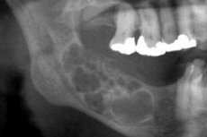
The radiographic picture of ameloblastoma is quite specific. The distinctive radiographic criterion is the different degrees of transparency of the cavity shadows. Cavities can have different levels of transparency: from low to high. The central part of the cyst is always highly transparent. In the cystic variant of ameloblastoma, one large cyst localized in the area of the mandibular angle and branch, or a polycystoma, can be detected. A large cyst is radiographically characterized by clear boundaries of the formation, often homogeneous bone rarefaction. In some cases, an impacted tooth is projected onto the cystic cavity, but its crown is located outside with different dental arrangements. An X-ray of a polycystoma demonstrates the presence of several cysts of different diameters, mutually adjacent (like "soap bubbles"). The formations have a clear rounded configuration, sometimes with uneven contours. They may contain an impacted tooth. [ 20 ]
Solid ameloblastoma is identified on radiographs by uneven bone rarefaction with relatively clear boundaries. In some patients, barely distinguishable cystic cavities are found against the background of rarefaction, which often indicate a transitional period of the neoplasm from solid to cystic ameloblastoma.
Differential diagnosis
Ameloblastoma should be differentiated from the following pathologies:
- osteoblastoclastoma;
- odontogenic cysts;
- fibrous osteodysplasia;
- sarcoma;
- chronic osteomyelitis (with a suppurating tumor).
If the tumor is located in the mandibular angle, it should be additionally distinguished from odontoma, hemangioma, cholesteatoma, fibroma, and eosinophilic granuloma.
Who to contact?
Treatment ameloblastomas
Ameloblastoma is cured only by surgery, namely, by removing the jaw tissue damaged by the tumor. The extent of the intervention is determined by the location and stage of the pathological process. The earlier the operation is performed, the fewer structures have to be removed. If the tumor has reached a large size and has spread to a predominant part of the bone, it may be necessary to remove part of the jaw and even the entire row of teeth. Since the operation is performed in the area of the face, where the aesthetic factor is especially important, the intervention is completed with reconstructive correction of the removed tissues and organs - that is, the elimination of the visible cosmetic defect. [ 21 ]
After resection of the tumor focus, drug therapy is started, aimed at preventing postoperative complications and recurrence of the pathology.
Antibiotics after surgery are prescribed by the surgeon. Amoxiclav is often the drug of choice due to its effectiveness, minimal contraindications and side effects. The medications are taken strictly according to the scheme described by the doctor.
If pain occurs, take analgesics and anti-inflammatory drugs (for example, Nimesulide), as well as vitamin supplements to support the immune system.
Chlorhexidine, furacilin solution, and Miramistin are usually used to rinse the mouth.
During the rehabilitation stage, it is important to follow a special diet. Food should be soft (optimally liquid), with a comfortable temperature. You should exclude hot spices, salt and sugar, soda, alcoholic beverages, and raw plant foods from your diet. [ 22 ]
Medicines
When choosing medications, it is necessary to take into account contraindications, the degree of toxicity of the drugs, possible side effects, the rate of penetration into soft tissues and the period of elimination from the body. [ 23 ] The following medications may be prescribed:
- Ibuprofen - take one tablet three times a day for three days. Longer use may negatively affect the digestive system.
- Ketanov - taken orally once or repeatedly, depending on the severity of pain, 10 mg per dose, up to 3-4 times a day. The duration of treatment is no more than five days, which helps to avoid erosive and ulcerative lesions of the gastrointestinal tract.
- Solpadeine - used to relieve severe pain, 1-2 tablets three times a day, keeping an interval of at least 4 hours between doses. The drug should not be taken for more than five days. With prolonged use, abdominal pain, anemia, sleep disorders, tachycardia are possible.
- Cetrin - to relieve swelling, take 1 tablet daily with water. The drug is usually well tolerated, only sometimes it can cause digestive discomfort, headache, drowsiness, dry mouth.
- Amoxiclav - in the postoperative period, 500 mg is prescribed 2-3 times a day, for a course of up to 10 days. Possible side effects: dyspepsia, headache, convulsions, allergic reactions.
- Tsifran (ciprofloxacin) – prescribed as part of antibiotic therapy in individual dosages. Possible side effects include nausea, diarrhea, and allergic reactions.
- Lincomycin is a lincosamide antibiotic taken 500 mg three times daily. Treatment may be accompanied by nausea, abdominal pain, reversible leukopenia, and tinnitus. Such side effects resolve on their own after the course of treatment.
Physiotherapy treatment
Physiotherapy can be used after surgical resection of ameloblastoma to speed up tissue recovery. Good results are provided by:
- electrical action of ultra-high frequencies in an oligothermic or athermic dose, lasting 10 minutes, six procedures per treatment course;
- fluctuation lasting 10 minutes, in the amount of six procedures (three daily, and the rest once every two days);
- infrared laser with a treatment duration of 15-20 minutes, daily, in the amount of 4 procedures;
- magnetolaser treatment with a wavelength of 0.88 µm, a total power of 10 mW, magnetic induction from 25 to 40 mT, with a duration of action of 4 minutes and a course of eight sessions.
If there are seals and cicatricial changes in the area of the operation, then ultrasound treatment is indicated in continuous mode, with a session duration of up to 8 minutes and a head area of 1 cm². The treatment course consists of 8-10 sessions.
Herbal treatment
How can herbs help with ameloblastoma? Some plants can relieve pain and stimulate the immune system, thereby accelerating tissue regeneration. Other benefits of herbal medicine are also known:
- herbs may have antitumor effects;
- many plants maintain acid-base balance;
- herbal preparations are well absorbed even by a weakened organism at any stage of the pathology;
- Herbs improve the body's adaptation to new living conditions and facilitate the course of the postoperative period.
Medicinal plants can be used both dried and freshly picked. They are used to make infusions and decoctions. The following types of herbs are relevant for ameloblastoma:
- Catharanthus is a semi-shrub with antitumor activity. To prepare the tincture, take 2 tbsp. of twigs and leaves of the plant, pour 250 ml of vodka, keep in a dark place for 10 days, filter. Take 5 drops half an hour before meals, increasing the dosage daily, bringing it to 10 drops per day. Duration of treatment is 3 months. Caution: the plant is poisonous!
- Marshmallow is a well-known expectorant and anti-inflammatory plant, which is no less effective in various tumor processes. One tablespoon of crushed rhizome is poured into a thermos with 200 ml of boiling water, kept for 15 minutes, poured into a cup and cooled at room temperature for 45 minutes, then filtered. Take orally three times a day after meals, 50-100 ml, for 2-3 weeks.
- Sweet flag - the rhizome of this plant contains a terpenoid that has an analgesic and restorative effect. Prepare an infusion of 1 tbsp. of crushed root per 200 ml of boiling water. Take 50 ml per day (divide into two doses).
- Barberry - contains an alkaloid, which is successfully used to treat even malignant tumors. The roots and young shoots of barberry (20 g) are poured with 400 ml of boiling water, boiled for 15 minutes, then infused for about 3-4 hours. Filter and bring the volume to 500 ml with boiled water. Drink 50 ml 4 times a day.
- Immortelle – excellent for relieving spasms and eliminating pain after surgery. To prepare the infusion, take 3 tbsp. of the crushed plant, pour 200 ml of boiling water, leave for 40 minutes, filter. Bring the volume to 200 ml with boiled water. Take 50 ml three times a day half an hour before meals for a month.
- Burdock root – has an antitumor effect. Taken orally as a decoction (10 g per 200 ml of water), 100 ml twice a day, for a month.
- Sedum - a decoction and infusion of this herb improves metabolism, tones, eliminates pain and stops the inflammatory process. Prepare an infusion of 200 ml of boiling water and 50 g of dry crushed leaves of the plant. Drink 50-60 ml daily.
- Thistle - prevents the development of tumor relapse. The infusion is prepared at the rate of 1 tbsp. of leaves per 200 ml of boiling water. Take the remedy 100 ml 3 times a day.
- Calendula - promotes the resorption of pathological foci, blood purification, and wound healing. Take the pharmacy tincture 20 drops 15 minutes before meals (with water) three times a day for a month.
The use of medicinal plants must be approved by the attending physician. They should never be used as a substitute for traditional treatment. [ 24 ]
Surgical treatment
Treatment consists of surgical removal of ameloblastoma. In case of purulent inflammatory process, the surgeon performs sanitization of the oral cavity. The neoplasm is enucleated, the walls are washed with phenol: this is necessary to start necrotic processes in tumor elements and slow down their development. If the operation is performed in the mandibular region, then bone grafting and dental prosthetics with constant wearing of an orthopedic device are additionally performed. Upon completion of the operation, the cavity is not sutured to reduce the risk of recurrent tumor development. Instead of applying a suture, tamponade is used, which promotes epithelialization of the cavity walls. [ 25 ]
In complex chronic cases, partial jaw disarticulation is performed (surgical twisting of the jaw along the border of the joint space, which does not require bone sawing). Instead of the removed part of the jaw, a bone plate is implanted using a special orthopedic device.
If removal of ameloblastoma is impossible for some reason, or if the tumor becomes malignant, radiation therapy is prescribed. [ 26 ]
After surgery, patients undergoing surgery are prescribed a course of antibiotics and are given the basics of postoperative nutrition. For several weeks, the patient should not eat hard or coarse foods, and after each meal, the mouth should be rinsed with a special solution. [ 27 ]
Removal of ameloblastoma is carried out as follows:
- If the neoplasm is localized in the bone mass, then a partial mandibular resection is performed.
- If the ameloblastoma is large and extends to the edge of the lower jaw, then a through mandibular resection is performed. If the branch has severe damage and the condylar process is affected, this is an indication for exarticulation of the lower jaw and the neoplasm to the borders of healthy tissue.
- To prevent recurrent tumor growth, the surgeon must have an understanding of and adhere to the principles of ablastics and antiblastics.
The patient is treated in hospital for about 2 weeks, after which he is transferred to outpatient observation with a mandatory visit to the doctor:
- during the first year after surgery – every three months;
- over the next three years – once every six months;
- then annually.
Prevention
In order to prevent complications in the form of inflammatory processes, pathological fractures and malignancy at the preoperative stage, it is necessary to detect ameloblastoma as early as possible. For all patients without exception, complex treatment with the use of symptomatic drugs and antibiotic therapy is recommended.
To prevent bleeding during the postoperative recovery stage, it is necessary to monitor the quality of blood clotting and blood pressure indicators.
Prevention of late adverse effects is closely related to qualified diagnostics, preliminary stereolithographic modeling. Radical intervention with subsequent bone plastic surgery, with the installation of endoprostheses and zealous implants, contour plastic surgery, and transplant microvascular measures is considered optimal.
Forecast
Ameloblastoma is often diagnosed at late stages of growth, which is due to the insufficiently pronounced symptoms of the disease and its small spread. The main treatment option for the tumor is its immediate removal with subsequent reconstruction (if possible).
The basic factor for a favorable prognosis is early diagnosis of the disease and timely qualified treatment, including surgical removal, chemical or electrical coagulation, radiation therapy, or a combination of surgery and radiation.
The further outcome of postoperative recovery depends on the volume and nature of the treatment performed, including surgery. For example, radical removal of the lower jaw entails the appearance of significant cosmetic defects, as well as impairment of speech and chewing function. [ 28 ]
The main point of rehabilitation of patients who have undergone radical interventions is considered to be correction of jaw function. For this purpose, primary or delayed bone plastic surgery is performed with subsequent dental prosthetics. The scope of such an operation is determined by a maxillofacial surgeon.
At present, methods of individual dental prosthetics after ameloblastoma has been removed from a patient have not been sufficiently developed, despite the fact that restoration of facial configuration and jaw functionality is an important point of social and medical rehabilitation.