Medical expert of the article

New publications
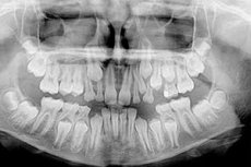
Orthopantomography - panoramic radiograph of the maxillofacial region
Last reviewed: 06.07.2025

All iLive content is medically reviewed or fact checked to ensure as much factual accuracy as possible.
We have strict sourcing guidelines and only link to reputable media sites, academic research institutions and, whenever possible, medically peer reviewed studies. Note that the numbers in parentheses ([1], [2], etc.) are clickable links to these studies.
If you feel that any of our content is inaccurate, out-of-date, or otherwise questionable, please select it and press Ctrl + Enter.
 [
[ Indications for the procedure
In most cases, this diagnostic method is used by dentists (for treatment, removal and prosthetics of teeth) and maxillofacial surgery specialists.
Dentists prescribe orthopantomogram of teeth for the purpose of visualization of the entire dental system of patients, providing objective data for assessing its condition. And this ensures the correctness of the diagnosis and the choice of the optimal tactics for treating the roots of teeth and their canals, as well as such dental diseases as root cyst, periodontitis, periodontosis, etc.
In prosthetics, an orthopantomogram of the jaw or a panoramic x-ray of the jaw allows one to clearly determine the degree of periodontal bone loss with an incomplete row of teeth and choose the most acceptable way of its restoration (using removable dental structures, dentures or dental implants).
Orthopantomogram of a child may be prescribed by orthodontists during examination of dystopia and retention (impaired eruption) of teeth, as well as defects in occlusion of dental arches (malocclusion). Such an X-ray examination is necessary when there is traumatic damage to the jaw bones or in the presence of:
New formations of bone tissue of the facial skeleton and jaws (osteoblastoclastoma, osteoma, odontomas, etc.);
Dermoid or bone cyst, cystic form of ameloblastoma of the jaw;
Congenital defects of the facial skeleton, in particular, craniofacial dysostosis, osteodysplasia, dysraphic syndrome (cleft lip or palate).
Indications for orthopantomography also include diagnostics of:
- bone and joint lesions of the maxillofacial region of the skull, including dentoalveolar fractures and fractures of the lower jaw;
- dysfunction of the temporomandibular joints;
- osteomyelitis of the mandible and condylar processes;
- salivary gland stones (sialolithiasis);
- osteosarcoma, osteoradionecrosis and radiation osteomyelitis of the jaw;
- ameloblastomas;
- ankylosis of the temporomandibular joint;
- maxillofacial dermoid cysts (teratomas);
- maxillary sinus cysts;
- calcifications and atherosis of the carotid artery.
A dental panoramic radiograph can also be used in the diagnosis of ENT diseases, since the device for this type of radiography captures the frontal sinuses, nasal cavity, part of the pharynx and neck.
Technique orthopantomograms
Preparation for orthopantomographic examination consists of the patient having to remove all metal costume jewelry and jewellery and put on a protective lead vest-apron covering the body, including the thyroid gland area and neck. The patient also bites a small plate connected to the device (in our clinics, German Orthophos XG devices of various modifications are most often used). It is important to remain completely still during the scanning (approximately 18-35 seconds).
Computer orthopantomogram (digital) saves images as files in the patient database (archive) – with unlimited possibility of their use both for comparison of treatment results and for modeling various maxillofacial surgeries.
Normal performance
The interpretation of an orthopantomogram, which notes the normal indicators of the dental structures and describes all anatomical deviations and morphological disorders, is carried out by radiologists who specialize in dentistry and maxillofacial pathologies.
For example, a benign bone tumor of the jaw, osteoma, will look like a dense bone area on an orthopantomogram. A cyst looks like a rounded area of less dense bone tissue with clear boundaries.
Harm and radiation exposure during orthopantomogram
The standard radiation dose during an orthopantomogram is 0.01-0.04 mSv (10-40 μSv). There is no harm to the body or side effects - if all protective measures are observed - since the dose of a single direct ionizing effect on cells is low, and the standards existing in medical radiology allow a maximum radiation load of 1000 μSv over 12 months.
Orthopantomogram has the following advantages: full coverage of facial bones and teeth; speed of examination and its convenience for the patient; the possibility of use when opening of the mouth is limited (for example, due to spasm of the masticatory muscles).