Medical expert of the article

New publications
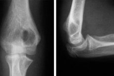
X-rays of the elbow joint.
Last reviewed: 03.07.2025

All iLive content is medically reviewed or fact checked to ensure as much factual accuracy as possible.
We have strict sourcing guidelines and only link to reputable media sites, academic research institutions and, whenever possible, medically peer reviewed studies. Note that the numbers in parentheses ([1], [2], etc.) are clickable links to these studies.
If you feel that any of our content is inaccurate, out-of-date, or otherwise questionable, please select it and press Ctrl + Enter.
Radiography is a diagnostic procedure with a solid track record, counting more than 120 years. But, despite the development of new modern methods of diagnosing various diseases, it has not lost its relevance to this day. X-ray equipment is available in almost all clinics, the examination procedure is simple to perform, and in terms of information content it is not much inferior to other methods. If the doctor suspects a joint pathology, X-ray will be the most basic method for diagnosing the problem. One of the most popular diagnostic procedures in traumatology is an X-ray of the elbow joint, a bone joint that can be damaged not only due to injuries, but also as a result of some pathological processes occurring in the body.
Indications for the procedure
Radiography is a method of diagnosing internal injuries, which is used in cases where the doctor has difficulty making a diagnosis based on external signs, or to clarify some details of tissue damage. X-rays make it possible to see through the body to detect pathological changes in muscles, bones, cartilage, etc., which are hidden from the human eye.
X-ray of the elbow joint, like other methods of examination using X-rays (ionizing radiation), is not a completely safe procedure, which can cause radiation burns and cell mutation, increase the risk of tumor development. However, the degree of danger of X-ray radiation depends on the duration and frequency of irradiation procedures. It is clear that such a diagnostic method as X-ray cannot be used purely out of curiosity. The doctor must have good reasons to refer a person for examinations.
As for elbow joint pathologies, the following are compelling reasons:
- unexplained pain in this area without external damage,
- swelling of soft tissues in the elbow area,
- change in tissue color (redness, bluish tint),
- local increase in temperature,
- complaints of limited arm movement at the elbow,
- elbow injuries accompanied by pain, redness of tissues and their swelling, both with a violation of the integrity of soft tissues and without visible damage.
As for traumatic injuries, X-rays can rule out or confirm fractures and dislocations that complicate treatment.
It should be said that X-rays are prescribed not only by traumatologists when there is a suspicion of a fracture of the humerus, ulna or radius or a dislocation of the forearm bones in the elbow area. If there was no injury, but suspicious changes in the color and structure of soft tissues are detected, pain in the elbow appears, limiting hand movements, first of all we go to the therapist, and he decides whether to send the person for an X-ray or offer a consultation with an orthopedist. All these doctors can give a referral for an X-ray of the elbow joint, if this is necessary to clarify the diagnosis.
Preparation
X-ray examination is one of the simplest diagnostic methods, if only because it requires virtually no preparation for the procedure. The only thing the doctor will ask is to free the arm below the shoulder from clothing, jewelry, and watches. No dietary or medication restrictions are required.
 [ 3 ]
[ 3 ]
Technique X-rays of the elbow joint.
An X-ray of the elbow joint is usually performed in a sitting position, but if necessary, diagnostics can also be performed in a lying position (for example, if the person is unconscious) or standing. The patient sits on a chair moved to a special table of the X-ray machine, sideways to it. The limb being examined is placed on the table in the position indicated by the doctor. The edge of the table should be slightly above the level of the armpits.
In order for the image to be clear, the limb must remain motionless during the examination. If the patient has difficulty holding the arm still, the limb is fixed on both sides with special bags filled with sand or other heavy material.
According to standards, joint radiography should be performed in 2 projections. A study in a direct projection requires maximally straightening the arm and placing it on the table so that the palm faces up. The arm at the elbow should be slightly raised.
For a lateral projection, the arm is bent at the elbow at a right angle and placed with the back of the hand facing up. The patient should sit at such a height that the shoulder and forearm are at the same level.
In some cases, examination is also required in another projection - axial, when the posterior part of the humerus and the olecranon are clearly visible. To conduct the examination, the arm must be fully bent at the elbow, as much as possible. On the table, the arm is resting on the humerus.
In all cases, the X-ray cassette is placed under the elbow. To protect the chest and body, patients are asked to wear a special apron made of X-ray-proof material.
Depending on the equipment used (film or digital), the results of the study can be obtained on a special film that requires preliminary development in a specially equipped room, or on a digital medium from which you can print the image on paper or view it on a computer monitor.
Digital radiography, which appeared much later than film radiography, is becoming increasingly popular, because it allows you to enlarge the image on the monitor, zoom in on its individual elements to examine the damage. And you can store the image on a disk for a long time without distortion. Perhaps in the future it will be needed for comparison if new injuries are received, or to evaluate the effectiveness of treatment. Such images can be archived and stored for a long time on the computer of a specialist doctor.
Contraindications to the procedure
An X-ray of the elbow joint, like any X-ray examination, is not considered a safe procedure due to some properties of ionizing radiation. And, despite the fact that it is done even to children if necessary, the procedure still has some limitations.
The main limitation is considered to be childhood. In theory, X-rays are allowed for children over 14 years old. We are not talking about possible pathologies, because usually the radiation dose and the duration of the procedure are adjusted in such a way that they cannot cause significant harm. It is just that the effect of ionizing radiation on a child's body is more pronounced and can affect the development of various systems of the child. And the younger the child, the more dangerous X-rays are for him. For example, in infants, many important body systems are in the formation stage, so cell mutations leading to disruption of their activity are more likely.
If necessary, X-rays are taken even on newborns, but all parts of the child's body except for the area being examined are covered with special protective equipment. Older children must have their chest, stomach, and pelvic area covered with a protective apron. The thyroid gland and eyes must also be protected from ionizing radiation.
If X-rays are so dangerous for small children, one can imagine what harm they can cause to an unborn baby with unformed vital systems. Exposure of a pregnant woman to radiation is fraught with the risk of giving birth to a child with various mutations and pathologies, so X-rays are contraindicated for expectant mothers.
X-rays of pregnant women can only be done according to strict indications, and the abdominal area must be protected with a lead apron that does not let X-rays through. Ideally, a protective apron should be used in all cases of X-rays to reduce the negative impact of radioactive radiation on the human body.
[ 4 ]
Normal performance
The elbow joint is a rather complex structure, including the humero-ulnar, humeroradial and proximal radio-ulnar joints. In order to carefully examine all these components and their parts, radiography is performed not in one, but in 2-3 projections. Accordingly, the results are deciphered according to all three components of the elbow joint, and not in general terms.
If the elbow joint X-ray is normal, the examination report will note that its general X-ray anatomical orientation does not differ from the usual one, and all the ratios of the sizes of bones and joints are standard. The components that form the joint are proportionate to each other, their size and shape are normal. In the image in direct projection, 3 joint spaces are clearly visible and distinguishable from each other, corresponding to 3 joints, united by the common name " elbow joint ":
- the humero-ulnar joint (the junction of the humeral block and the coronoid process of the ulna) is a simple block joint,
- the humeroradial joint (the place of articulation of the elevated part of the head of the humerus and the cavity of the head of the radius) is a simple ball-and-socket joint,
- The proximal (upper) radioulnar joint (the junction of the circumference of the radius and the radial cavity of the ulna) is a simple cylindrical joint.
The width of the joint spaces in the ball and socket joints should be the same and have a standard size.
In the anatomy of the human skeletal system, there are such concepts as epiphysis, diaphysis and metaphysis of the bone. The epiphysis of the bone is called the enlarged rounded end of the tubular bone (its head, including convex and concave parts), which forms the joint. The articular part of the epiphysis is covered with cartilage.
The diaphysis is nothing more than the central part of the tubular bone (its body). Between the epiphysis and the diaphysis is the metaphysis (in childhood and adolescence it is responsible for bone growth), adjacent to the cartilaginous epiphyseal plate, which in turn is articulated with the subchondral plate, which has many capillaries and nerve endings.
On an X-ray of a normal elbow joint, the cartilaginous tissue of the epiphyses of the bones (also called the endplate of the epiphysis or the cartilaginous growth plate) should have a smooth and clear outline. The subchondral part of the epiphysis should have its characteristic porous (spongy) structure.
The visible areas of the metaphysis should have a normal shape without thickenings, the structure of the bone tissue should correspond to the patient's age (ossification of the metaphysis occurs as a person ages and is completed at 18-25 years).
The visible areas of the diaphysis of the bones should also have a normal shape and structure without cracks, displacements, thickenings, or bends.
The elbow joint also has some soft tissue parts. These include the joint bag (joint capsule) and intra-articular ligaments. An X-ray of a normal joint does not reveal ossification in these parts (bone tissue on a black-and-white X-ray has a lighter shade). The soft tissues surrounding the joint should have the appropriate volume (mass), structure and shape, which indicates the absence of tumors and degenerative changes.
But so far we have talked about normal elbow joint X-ray indicators. Now let's try to understand what a doctor sees when a patient with one of the most popular elbow pathologies comes to him, because in most cases the result will not be as cloudless as we saw above. After all, it is not healthy people who seek medical help.
For example, a person goes to the doctor complaining of severe spontaneous pain in the elbow, which increases with arm movement and physical activity. At the same time, muscle strength weakens. Such symptoms may indicate elbow epicondylitis - an inflammatory-degenerative disease of the elbow tissues that affects the bones, periosteum, tendons, and is the result of constant overload of the elbow joint.
Symptoms of epicondylitis, which often affects people of certain professions, are similar to other pathologies (arthritis, bursitis, soft tissue bruises, cracks in the styloid process of the ulna or radius, epicondyle fracture, tunnel syndrome, etc.). Differential diagnostics help to differentiate one disease from another. However, it rarely relies on the results of an X-ray. At the onset of the disease, an X-ray can only exclude joint dislocations and bone cracks, but it is impossible to diagnose epicondylitis itself with its help.
But when the disease becomes chronic, characterized by degenerative changes in the joint tissues, an X-ray will help not only diagnose the disease, but also assess the degree of joint damage in order to determine the methods of treating the pathology.
X-ray signs of chronic epicondylitis of the elbow joint are foci of osteoporosis (destruction of bone tissue), bone growths (osteophytes) formed as a result of prolonged inflammation, compaction at the ends of tendons and in porous bone structures. Since bone structures transmit X-rays worse than soft tissues, there will be more light spots on the image than necessary, and in areas of osteoporosis, the color, on the contrary, will be closer to gray.
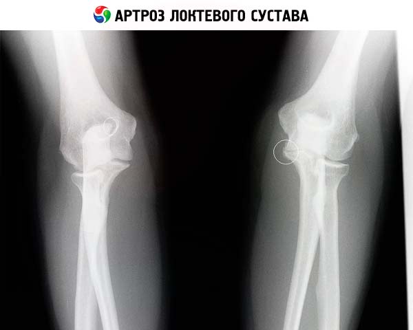
For example, in the case of a no less popular pathology called elbow arthrosis, the X-ray first of all shows a narrowing of the joint spaces, which makes it difficult to move the arm and bend it at the elbow. This can be seen by a too thin strip (up to its absence) in place of the joint space. The contours of the cartilaginous tissue in the joint area will also be changed.
As for another fairly common pathology, such as elbow dislocation, which occurs in both adults and children, X-rays are often not required. The symptoms of dislocation are quite clear: severe pain in the elbow joint, limitation of its mobility due to a strong increase in the intensity of the pain syndrome, swelling of the soft tissues in the affected area, a strong decrease in the sensitivity of the hand. In addition, the doctor cannot feel the pulse on the hand below the elbow, but the protruding end of the radius is usually well palpated.
Depending on the conditions under which the dislocation occurred (when falling on an arm extended or bent at the elbow), a posterior, lateral (the forearm bones in the joint are displaced backwards and inward or outward) or rarer anterior dislocation can be diagnosed.
The main X-ray signs of elbow dislocation:
- Lack of contact between the articular surfaces of the bones with a violation of their location relative to each other. In the joints, the cavity of one bone is filled with the convexity of the head of the other; in case of dislocation, the cavity is empty. Depending on how much the bones are displaced relative to each other, a complete or incomplete dislocation of the elbow is diagnosed. In the latter case, part of the head of one bone comes into contact with the cavity of the other bone.
- Dislocation of the axis of the dislocated bone. This sign is very relevant when X-raying the elbow joint in children, because the distal parts of the bones in a child are still in the process of ossification, so it is very difficult to assess the changes in the size of the gap between the bones (cartilage transmits X-rays almost as well as soft tissues, so they are practically invisible on the X-ray, and the joint gap should be understood as the distance between the ossified areas). However, with a lateral fracture, the degree of bone displacement is very difficult to assess, so it is necessary to take pictures in different projections.
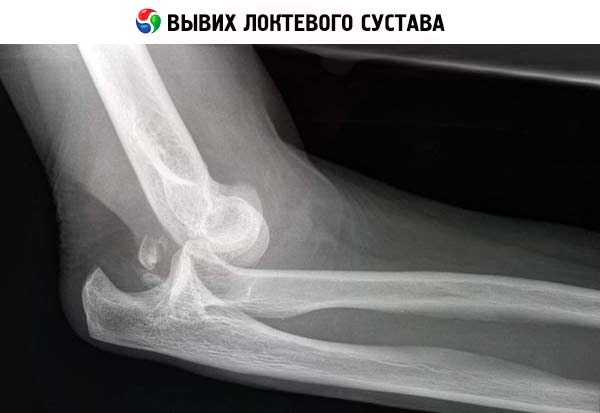
In one third of cases of traumatic elbow dislocations, small bone fragments are torn off at the point where the joint capsule and ligaments are attached to them. Small fragments are usually not dangerous and do not interfere with reducing the dislocation. But if we are talking, for example, about a torn medial epicondyle, which sometimes happens with an external elbow dislocation, sometimes it is necessary to resort to surgical intervention to remove the detached piece of bone (which does not allow the dislocated bone to be put back in place. On an X-ray, the fragment is visible as an area of abnormal lightening in shape and size corresponding to the formed notch on the damaged bone.
When an old dislocation is detected on the image, which was not reduced at the time, the picture may be as follows: osteoporosis or destruction of the distal sections of the displaced bones with a change in their shape and size, atrophy of soft and hard tissues in the joint area, formation of a new glenoid cavity (neoarthrosis). The presence of such signs and their severity depend on the "age" of the dislocation. After a recently reduced dislocation, no changes in the joint tissues are observed, unless we are talking about a torn piece of bone.
X-rays also help to identify pathological dislocations, which do not necessarily have to be preceded by trauma. A person may not even suspect such damage as a result of strong muscle tension or harmless trauma. Pathological dislocations are caused by inflammatory processes in the joint area with a constant accumulation of liquid contents there. This leads to stretching of the joint capsule and the bones in the joint can shift even with minor mechanical impact.
Other causes of pathological dislocations include osteoarthrosis, tumors at the articular ends of bones, congenital defects in bone structure, etc. But whatever the cause of the pathological dislocation, a person comes with pain and limited hand movement and does not associate them with a dislocation. X-ray diagnostics can give a clear picture of the pathology. It will also allow differentiating a bone dislocation from a fracture or chip, the symptoms of which are outwardly similar to each other.
Complications after the procedure
Let's say that X-rays are most dangerous in childhood, so they are prescribed as a last resort, when there is no possibility to resort to safer diagnostic methods: ultrasound examination (US) or magnetic resonance imaging (MRI). Computer tomography (CT) is not so safe in this regard and can have consequences similar to X-ray irradiation during radiography (the same frequencies are used).
What is dangerous about X-rays? Their radioactivity and ability to change the properties of cells, which results in disruption of organ functionality and active proliferative processes in them, ultimately leading to the development of tumor processes. We had the opportunity to observe a similar situation on a large scale after the explosion at the Chernobyl Nuclear Power Plant, the consequences of which are still echoing among its witnesses to this day.
But the situation with X-ray examination is somewhat different. We are talking about completely different doses of radiation. The dose of X-ray radiation is not much different from the dose of radiation we receive when flying on airplanes or going through an introscope at the airport, so there is no point in talking about possible complications. Many men, women and children use Aeroflot services several times a year, and this does not affect their health in any way. What can I say, some people live in areas with unfavorable radiation conditions, where radiation doses approach X-ray radiation.
It should be noted right away that not only the radiation dose, but also the duration of exposure to rays during radiography are strictly limited, so 1-3 images per year, and an X-ray of the elbow joint is unlikely to have to be done more often, will not be able to cause significant harm to the patient's body, but will help to identify dangerous pathologies and assess the effectiveness of the treatment. Even a child can have about 5-6 images per year without consequences.
But again, it is necessary to take into account the radiation background of the area where a person lives, and the frequency of using services that involve irradiation of the body. It is desirable that the total dose of radiation received by a person during the year from various sources does not exceed 3-4 millisieverts.
Reviews
An X-ray of the elbow joint is a fairly informative non-invasive procedure that can be performed in almost any clinic, since it does not require the purchase of expensive modern equipment (although modern X-ray machines are considered safer in terms of radiation).
With the help of X-rays, it is possible to examine degenerative-dystrophic processes in tissues located deep inside the body, penetrate deep into bone tissue to assess its structure and possible changes, identify fractures of various parts of the bone and congenital anomalies that predispose to injury with the slightest mechanical impact. And the doctor has the opportunity to see all this without surgical intervention, because soft tissues remain transparent to X-rays.
Another advantage of such examination is the absence of any special preparation for the procedure. A person does not need to limit themselves in food, liquids, medicines, prepare the skin, etc. And there is no specialized care after the procedure. After taking the results of the examination a quarter of an hour later, a person goes to the attending physician, who prescribes the appropriate treatment.
If a person is afraid of the dose of ionized radiation, he can drink a glass or two of homemade milk at home, which helps remove radiation from the body. The same is recommended for people who live or work in regions with an increased radioactive background, but the milk should not be local, but delivered from ecologically clean areas.
The harm from X-rays, according to doctors, is significantly less than its benefits. After all, even the reduction of non-specific dislocations should occur under its control. Not to mention the possibility of identifying hidden pathologies that a person might not even suspect for a long time.
Elbow injuries and degenerative changes in its joints are considered to be quite common pathologies, and X-ray of the elbow joint is considered to be a rather popular procedure. Yes, there are now safer methods for diagnosing bone pathologies, however, X-ray remains one of the most widely used and very inexpensive methods available to almost everyone.