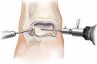
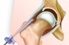
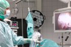
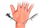
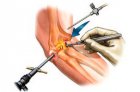
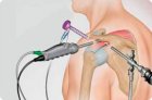
^

Health

Examination of the body

Color vision

The study of color vision can be informative in the clinical evaluation of hereditary dystrophies of the retina, when its disturbances occur before the visual acuity decreases and scotomas appear.

Visual evoked potentials

Visible evoked potentials (VEP) are recorded to diagnose lesions of the visual pathways, assessing their condition from the peripheral (retina) to the central sections (primary and secondary visual centers).

Electrooculography

Electrooculography - registration of the permanent potential of the eye with the help of skin electrodes applied to the area of the outer and inner margins of the lower eyelid.

Electroretinography

Electroretinography is a method for recording the total bioelectrical activity of all retinal neurons: negative a-wave - photoreceptors and positive b-wave - hyper- and depolarizing bipolar and Mueller cells.

Pachymetry of the cornea

Pachymetry is the measurement of the thickness of the cornea. It is an indirect sign of the integrity of the corneal endothelium. The largest thickness of the cornea is near the limbus (0.7-0.9 mm).

Soskob and biopsy of the cornea

Scraping of the cornea is performed with a Kimura spatula, a curved end of the needle (for the hypodermis), or a blade. After instillation of a local anesthetic without preservatives, the edges and the bottom of the lesion (usually ulcers) are gently and carefully scraped under the control of the slit lamp.

Keratometry of the cornea

Kerometry of the cornea is the measurement of the curvature of the axial meridians of the anterior surface of the cornea.

Assessment of the sensitivity of the cornea

Assessment of the sensitivity of the cornea in order to identify gross violations of the sensitivity of the cornea.

Confocal intravital microscopy of the cornea

Confocal microscopy of the cornea is one of the modern methods of investigation; allows to conduct intravital monitoring of the cornea state with visualization of tissues at the cellular and microstructural level.

Biomicroscopy of the cornea

It is essential to systematically identify clinical signs, determine the localization, depth and size of corneal damage.

Pages

You are reporting a typo in the following text:
Simply click the "Send typo report" button to complete the report. You can also include a comment.