Medical expert of the article

New publications
Elbow arthroscopy
Last reviewed: 04.07.2025

All iLive content is medically reviewed or fact checked to ensure as much factual accuracy as possible.
We have strict sourcing guidelines and only link to reputable media sites, academic research institutions and, whenever possible, medically peer reviewed studies. Note that the numbers in parentheses ([1], [2], etc.) are clickable links to these studies.
If you feel that any of our content is inaccurate, out-of-date, or otherwise questionable, please select it and press Ctrl + Enter.
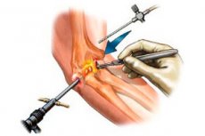
Recently, arthroscopy of the elbow joint has become widespread and introduced into clinical practice. In addition to purely diagnostic purposes (revision of intra-articular structures, biopsy of the synovial membrane and articular cartilage), various surgical manipulations are performed: removal of intra-articular bodies, sanitation of chondromalacia foci, arthrolysis, etc.
 [
[ Methodology for performing elbow arthroscopy
First, the elbow joint is marked with flexion to 90°: the lateral and medial epicondyles of the humerus, the head of the radial bone and all arthroscopic approaches used are marked.
Position of the patient
Supination position. The patient is positioned on his back, the arm is abducted at the shoulder joint to 90°. The distal part of the forearm and the hand are fixed in such a way that, if necessary, traction can be performed through a special suspension device with a block and counterweight attached to the operating table. In this case, the flexion at the elbow joint is maintained at an angle of about 90°.
Pronation position. The patient is in the prone position. The arm being examined hangs freely over the edge of the operating table. In this variant, a suspension system is not needed, the shoulder is abducted to 90°, and the elbow joint spontaneously sets a flexion angle of 90°. A short support with a roller is placed under the shoulder joint and the upper third of the shoulder.
A pneumatic tourniquet is applied to the upper third of the shoulder. Maximum pressure is 250 mm Hg.
At the first stage, the elbow joint cavity is filled to the maximum with a saline solution, which allows the nervous and vascular structures to be displaced forward and eliminates the possibility of their damage. The joint is filled through a direct lateral approach, in which a permanent outflow cannula is installed. Topographically, this approach is located in the center of the so-called Smith triangle formed by the middle of the head of the radius, the apex of the olecranon, and the lateral epicondyle of the humerus. The needle is inserted perpendicular to the skin surface through the muscles and joint capsule. Usually, the volume of the joint cavity is 15-25 ml. A sign that the joint is filled to the maximum is the outflow of fluid from the needle under pressure. The recommended pressure in the joint cavity is up to 30 mm Hg. At higher pressure, overstretching of the radial nerve may occur along with overstretching of the capsule.
Three main approaches are most often used in elbow arthroscopy: anterolateral, anteromedial, and posterolateral. Other approaches are considered additional and are used as needed. "Blind manipulation" of instruments in the joint cavity is unacceptable: this can lead to damage to the vascular-nerve bundle and/or articular cartilage even with maximum filling of the joint cavity.
Diagnostic arthroscopy of the elbow joint begins with the anterior section. This is due to the fact that maximum expansion of the joint cavity is possible only under the condition of maintaining the tightness of the joint capsule, and when performing a posterior approach, this condition is no longer met - accordingly, there is no maximum filling and movement of neurovascular structures forward.
Anterolateral approach. According to JR Andrews (1985), this approach is located 3 cm distal and 1 cm anterior to the lateral epicondyle. In this case, when inserted, the trocar passes ventral to the head of the radius through the short radial extensor carpi, just 1 cm from the radial nerve located anteriorly. WG Carson (1991) defines the point for this approach as 3 cm distal and 2 cm anterior to the lateral epicondyle, thereby getting even closer to the radial nerve. In an experiment on cadaveric specimens, we worked out what we believe to be the optimal point for this approach: it is located 1 cm distal and 1 cm anterior to the lateral epicondyle. A 0.5 cm long skin incision is made in the longitudinal direction. The arthroscope sheath with a blunt trocar is inserted strictly in the direction of the coronoid process. The trajectory passes straight, in front of the head of the radius, through the short radial extensor and 1 cm from the radial nerve. The arthroscope is inserted with pronation of the forearm, which reduces the risk of damage to the deep branch of the radial nerve.
First, the medial part of the joint capsule is examined.
In some cases, wrinkling and scarring of the medial part of the joint capsule can be noted. In case of hypertrophy of synovial villi, which complicates examination of the joint, shaving of the synovial membrane is performed.
The arthroscope is then moved from the medial to the middle and then to the lateral part of the joint. The humeral trochlea, coronoid process, head of the humeral condyle and head of the radius are examined sequentially. When examining these structures, attention is paid to the condition of the cartilaginous cover, the presence of foci of chondromalacia, their prevalence, the depth of the lesion of the cartilaginous plate, the presence of osteophytes of the coronoid process, its deformation and compliance with the humeral trochlea during flexion and extension. The head of the humeral condyle is examined from the front, the head of the radius - during rotational movements of the forearm, which makes it possible to examine approximately three quarters of its surface.
The next step is to determine the anteromedial approach, located 2 cm distal and 2 cm anterior to the medial epicondyle. The trocar trajectory in this case passes very close to the main vascular-nerve bundle. The studies of Lynch et al. (1996), as well as our observations, showed that when the joint is not filled with saline, the arthroscope passes only 6 mm from the median nerve and the nearby brachial artery, the bifurcation of which is approximately at the level of the neck of the radius. When the joint is filled, the main vascular-nerve bundle shifts 8-10 mm anteriorly. In addition, when passing the trocar, it is necessary to straighten the patient's arm to 110-120°. This is due to the fact that there is a so-called mobile ulnar nerve, which, when the elbow joint is flexed, can move to the medial condyle of the humerus and, accordingly, can end up in the zone of passage of the trocar or other arthroscopic instruments. This access is considered instrumental.
There is a second method of establishing the anteromedial approach. In this case, the arthroscope, inserted through the anterolateral approach, is advanced into the lower medial part of the joint. Then the arthroscope is replaced with a long trocar, which rests against the medial wall of the joint, and an incision is made on the skin from the outside in the area of the protruding end of the trocar. In our opinion, the second method has advantages, since there is no risk of damaging the articular cartilage when inserting the trocar. In addition, the point selected in the joint cavity under visual control is maximally distant from the anterior surface of the joint and, therefore, from the vascular-nerve bundle.
During arthroscopy, inversion, i.e. rearrangement of the arthroscope and instruments, is possible, since the best visualization of the synovial membrane of the lateral part of the joint, the head of the humeral condyle and the head of the radius is achieved from the anteromedial approach.
The main diagnostic approach for the posterior part of the joint is considered to be the posterolateral approach, localized 3 cm proximal to the apex of the olecranon, immediately behind the lateral edge of the triceps tendon. The branches of the posterior cutaneous nerve of the forearm and the lateral cutaneous nerve of the arm pass through the access zone. To prevent their damage, it is necessary to exclude the use of a sharp trocar when performing the access.
The second method of establishing the posterolateral approach is along the joint space between the direct posterior and mid-lateral approaches. In this case, the arthroscope passes into the olecranon fossa from the bottom up, which has its advantages for the review. The instrumental approach will then be the direct posterior one. The posterolateral approach allows visualization of the olecranon fossa, the apex of the olecranon, and the posterolateral side of the humero-ulnar joint. During the examination, it is necessary to perform flexion-extension movements in the joint, which allows for a more complete examination of this area.
The direct posterior approach is just lateral to the midline through the olecranon. The trocar is passed directly through the triceps tendon toward the center of the olecranon fossa. This approach is used to insert the arthroscope, while instruments are passed through the posterolateral approach.
After performing arthroscopy, sutures are applied to the skin wounds. Immobilization of the limb is indicated - on a sling bandage. The next day, active movements in the elbow joint begin.
Contraindications to elbow arthroscopy
Contraindications to arthroscopy are in the following cases:
- presence of general and local infection;
- deforming arthrosis of grades III - IV with significant narrowing of the joint space and deformation of the articular ends;
- severe contractures of the elbow joint with a decrease in the volume of the joint cavity.
Mistakes and complications during elbow arthroscopy
According to the literature, the most serious complications during elbow arthroscopy are neurovascular. GJ Linch et al. (1986) reported the results of 21 elbow arthroscopies. One patient had short-term paresis of the radial nerve, associated, in the author's opinion, with overstretching of the joint cavity, another had short-term paresis of the median nerve, caused by the action of a local anesthetic, and a formed neuroma of the medial cutaneous nerve of the forearm. JR Andrews and WG Carson (1985) also reported temporary paresis of the median nerve. With sharp and rough manipulations with arthroscopic instruments in the joint cavity, damage to the articular cartilage is possible.
In conclusion, it should be noted that arthroscopy of the elbow joint is a promising method of examination and treatment. Low trauma, maximum diagnostic value, as well as the possibility of combining arthroscopy with open surgical interventions make it possible to significantly increase the effectiveness of treatment of very complex intra-articular pathology of the elbow joint.