Medical expert of the article

New publications
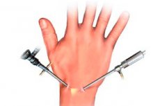
Carpal tunnel arthroscopy
Last reviewed: 04.07.2025

All iLive content is medically reviewed or fact checked to ensure as much factual accuracy as possible.
We have strict sourcing guidelines and only link to reputable media sites, academic research institutions and, whenever possible, medically peer reviewed studies. Note that the numbers in parentheses ([1], [2], etc.) are clickable links to these studies.
If you feel that any of our content is inaccurate, out-of-date, or otherwise questionable, please select it and press Ctrl + Enter.
The wrist joint is a complex of joints that connect the hand to the forearm. The wrist joint includes the radiocarpal, distal radioulnar, carpal, intermetacarpal, carpometacarpal and intercarpal joints. The wrist joint is small in size and is formed by 8 carpal bones, the radius and ulna, and a triangular fibrocartilaginous complex (cartilaginous articular disc).
Wrist joint injuries are varied and may arise from traumatic, infectious-inflammatory, degenerative and congenital causes. Among all musculoskeletal injuries, wrist joint injuries and diseases account for 4 to 6%.
The complexity of the anatomical structure, the variety of movements and the high functional demands placed on the wrist joint necessitate the most precise and careful performance of surgical manipulations on the wrist joint when it is damaged. In this regard, arthroscopic surgery is becoming increasingly important.
Arthroscopy allows direct visualization of all intra-articular structures of the wrist joint: articular surfaces, synovial membrane, ligaments of the wrist bones, etc.
In acute injuries of the capsular-ligamentous apparatus, arthroscopy is recommended in cases where after a period of fixation necessary for normalization of the joint condition and subsequent rehabilitation treatment, the condition does not improve. Previously, in such situations, in order to clarify the nature of the intra-articular injuries received, doctors were forced to use ultrasound, MRI, and contrast arthrography. However, the effectiveness of arthroscopy of the wrist joint has significantly changed this situation: arthroscopy, along with the indicated diagnostic methods, has made it possible not only to detect, but also to simultaneously correct intra-articular deviations. In some cases (according to various authors, up to 75%), arthroscopy allows us to identify damage to the triquetral fibrocartilaginous complex, lunate-triquetral and lunate-scaphoid instability, chondromalacia of the articular surfaces and joint fibrosis, while in most cases, when conducting a special radiation examination (ultrasound, MRI), pathological changes are not detected.
 [
[ Indications for wrist arthroscopy
Currently, it is difficult to formulate clear criteria for the need for surgical treatment. This is due to the fact that the indications for arthroscopy of the wrist joint are constantly expanding. They are determined based on the assessment of the integrity of the ligamentous apparatus: in the presence of ligament damage, the degree of rupture is assessed, as well as the presence of instability associated with this damage. Of great importance are the presence and degree of damage to the triangular fibrocartilaginous complex, identified cartilaginous defects in the wrist and intercarpal joints, and chronic wrist pain of unknown etiology.
Thanks to arthroscopy, it has become possible to perform the following treatment and diagnostic procedures in a minimally invasive and low-traumatic manner.
- Control of fragment reposition during extrafocal or minimally invasive osteosynthesis of intra-articular fractures of the wrist bones.
- Instabilities of interosseous joints (suturing, vaporization, radiofrequency ablation of ligaments).
- Damage to the triangular fibrocartilaginous complex (suturing, resection or debridement).
- Arthroscopic synovectomy.
- Detection and removal of intra-articular bodies.
- Ganglionectomy.
- Sanitation and lavage of the wrist joint.
- Carpal tunnel syndrome.
Technique of wrist arthroscopy surgery
The space available for arthroscopic manipulations in the wrist is significantly smaller than in larger joints. Wrist arthroscopy requires smaller diameter instruments (2.7-2.9 mm with a viewing angle of 30 and 70°). Precise placement and correct selection of instruments make it possible to ensure normal visualization of all structures and perform manipulations on all parts of the wrist joint.
To artificially enlarge the joint cavity during arthroscopy, it is necessary to apply traction to the wrist. The degree of traction varies and depends on the tasks being performed. There are several traction techniques.
- A specially designed universal traction system is applied.
- A preliminary application of an external fixation device is carried out, with the help of which distraction is performed.
- The assistant applies manual traction to the wrist or the first finger.
Knowledge of normal joint anatomy and accurate placement of arthroscopic portals are of the utmost importance for successful wrist arthroscopy. Inappropriate portal placement may not only interfere with the procedure but may also result in additional damage to intra-articular or periarticular structures.
Portal 3-4 is standardly used for visualization; 4-5 and 6-R are the main working portals for performing various manipulations. The outflow is established through portal 6-U.
Complications of wrist arthroscopy
If the surgical technique is performed correctly, complications from wrist arthroscopy are extremely rare. They can be prevented by following these guidelines.
- The surgeon must be absolutely precisely oriented in the anatomical structure of the joint, must be familiar with the anatomical landmarks and the location of the arthroscopic portals.
- It is necessary to observe the correct positioning and direction of the portals. The instrumentation should always be directed along the portals so that the instrument does not end up in the soft tissues outside the joint instead of in the joint cavity.
- To avoid damage to intra-articular structures, it is important to use blunt trocars and perform manipulations only with clear visualization of the working surface of the instruments inside the joint.
- A well-established drainage system prevents fluid from entering soft tissues.
- The use of saline solution promotes rapid absorption of fluid into soft tissues, thus reducing the risk of compartment syndrome.