Medical expert of the article

New publications
Color vision
Last reviewed: 06.07.2025

All iLive content is medically reviewed or fact checked to ensure as much factual accuracy as possible.
We have strict sourcing guidelines and only link to reputable media sites, academic research institutions and, whenever possible, medically peer reviewed studies. Note that the numbers in parentheses ([1], [2], etc.) are clickable links to these studies.
If you feel that any of our content is inaccurate, out-of-date, or otherwise questionable, please select it and press Ctrl + Enter.
Color vision testing may be informative in the clinical evaluation of hereditary retinal dystrophies when color vision impairment occurs before visual acuity declines and scotomas appear.
 [
[ Basic principles of color vision research
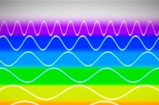
Color vision is provided by the functioning of 3 types of cones, each of which has its own maximum spectral sensitivity: blue (tritan) - 414-424 nm, green (deuteran) - 522-539 nm and red (prota) - 549-570 nm. All 3 types are required for normal perception of the visible spectrum. Color anomaly can concern each cone pigment: color weakness (for example, protanomaly - weakness of perception of red) or absence of color perception (for example, protanopia - absence of perception of red). In trichromacy, all 3 types are functionally active (but not necessarily functionally complete), while the absence of perception in the spectrum by one of the cone types is called dichromacy, and by two - monochromacy. Most people with congenital color vision disorders are anomalous trichromats with a violation of the proportion of the contribution of one or another part of the spectrum to their color perception. Impaired perception of red color due to functional deficiency of red cones is called protanomaly, green cones - deuteranomaly, blue cones - tritanomaly.
Acquired diseases of the macular region are characterized by more pronounced defects revealed by blue-yellow perimetry, and diseases of the optic nerve - by red-green.
Methods of studying color vision
- Ishihara charts are used to study people with congenital defects in red and green color perception. The 16 charts contain balls that form shapes or numbers that the person being tested must recognize. A person suffering from a color anomaly is unable to distinguish all the figures, and the inability to name the test object (with sufficient visual acuity) indicates simulation.
- The City University test includes 10 tables, each consisting of one central color and four peripheral colors. The subject must select the peripheral color that is most comparable to the central color.
- The Hardy-Rand-Rittler test is similar to the Ishihara charts but is sensitive to all three types of birth defects.
- The Farnsworth-Munsell 100-hue test is informative for congenital and acquired color vision disorders, but is rarely used in practice. Despite its name, it consists of 85-hue chips in 4 sections. The outer chips are fixed, the rest can be mixed by the researcher.
- the subject is asked to lay out the mixed chips in the correct order;
- the box is closed, turned over and the numbers inside the chips are evaluated;
- the data are marked in a simple cumulative manner on a circular map;
- Each form of dichromacy is characterized by insufficient color perception in its own meridian.
- The Farnsworth 15-Hue Test is similar to the Farnsworth-Munsell test, but consists of 15 chips.
For more information on testing color perception, read this article and Rabkin's tables in this one.