Medical expert of the article

New publications
Test of color perception and color vision: how to pass
Last reviewed: 04.07.2025

All iLive content is medically reviewed or fact checked to ensure as much factual accuracy as possible.
We have strict sourcing guidelines and only link to reputable media sites, academic research institutions and, whenever possible, medically peer reviewed studies. Note that the numbers in parentheses ([1], [2], etc.) are clickable links to these studies.
If you feel that any of our content is inaccurate, out-of-date, or otherwise questionable, please select it and press Ctrl + Enter.
Man is one of the few living creatures who are lucky enough to see the world in all its diversity of colors. But, alas, not everyone sees the surrounding objects the same way. There is a small percentage of people, mainly men, whose perception of colors is somewhat different from the majority. Such people are called colorblind, and if in life their vision peculiarity practically does not bother them (many may not suspect about the deviation for a long time), then when choosing a procession and passing a medical commission, some problems may arise. The thing is that areas of activity associated with a risk to the lives of others require correct recognition of colors. We are talking about such professions as a doctor, a driver of motor transport, a machinist, a pilot, a sailor, where one of the elements of professional selection is a color perception test. Problems with the implementation of labor activities may arise for colorblind people in the textile industry, landscape and interior design, work with chemical reagents, etc.
Color vision disorders
Scientists started talking about the fact that not all people can see the same object in the same color back in the late 18th century, when John Dalton described in his works the history of his family, in which he and his two brothers had a disorder of red color perception. He himself learned about this vision feature only in adulthood. It is worth saying that D. Dalton did distinguish colors, and did not see objects in black and white. It is just that his perception of colors was somewhat different from the traditional one.
Since then, the pathology of vision, in which a person sees colors differently, has been called color blindness. Many of us are accustomed to considering color blind people who perceive only black and white tones. This is not entirely correct, because color blindness is a generalized concept, within which several groups of people with different color perception are distinguished.
A person distinguishes colors thanks to the special structure of his visual organ, in the central part of the retina of which there are receptors sensitive to light of a certain wavelength. These receptors are usually called cones. The eye of a healthy person contains 3 groups of cones with a certain protein pigment sensitive to red (up to 570 nm), green (up to 544 nm) or blue (up to 443 nm) color.
If a person has all 3 types of cones in his eyes in sufficient quantity, then he sees the world naturally, without distortion of the existing colors. People with normal vision, according to scientific terminology, are called trichromats. Their vision distinguishes 3 primary colors and additional colors formed by mixing the primary shades.
If a person lacks cones of one of the colors (green, blue, red), the image is distorted, and what we see, for example, as blue, he may see as red or yellow. These people are called dichromats.
Among dichromats, there is already a division into groups depending on what color cones are missing in the eyes of patients. People with no receptors sensitive to green are called deuteranopes. Those who lack blue pigment are called tritanopes. If there are no cones with red pigment in the organs of vision, we are talking about protanopia.
So far we have been talking about the absence of cones of a certain pigment. But a certain part of people have all three types of cones, nevertheless, their color perception is somewhat different from the traditional one. The reason for this condition is a deficiency of cones of one of the pigments (they are present, but in insufficient quantities). In this case, we are not talking about color blindness in the literal sense of the word, but about anomalous trichromacy, in which the perception of colors is weakened. With a deficiency of red cones, we speak of protanomaly, with a lack of blue or green - respectively, about tritanomaly and deuteranomaly.
In the absence of color-sensitive cones, a person cannot distinguish colors and sees only different shades of black and white (achromatopsia). An identical picture is formed in those people whose visual organ contains cones of only one color (cone monochromacy). In this case, a person can see only shades of green, red or blue, depending on the type of cones present. Both groups of people are united by the common name of monochromats.
This pathology is rare, however, it has the most negative impact on a person's life, severely limiting his or her professional choice. Monochromats have problems not only with choosing a profession, but also with obtaining a driving license, because they naturally have difficulties recognizing the signal colors of traffic lights.
Most often, people with a violation of color perception of red and green colors are encountered. According to statistics, this pathology is diagnosed in 8 men out of 100. Among women, color blindness is considered a rare phenomenon (1 out of 200).
People with impaired perception cannot be blamed for their pathology, because in most cases it is congenital (genetic mutation of the X chromosome or changes in chromosome 7). However, there is a certain percentage of people in whom the pathology is considered acquired and affects mainly one eye. In this case, color vision impairment can be temporary or permanent, and is associated with age-related changes (clouding of the lens in the elderly), medication (side effects), and some eye injuries.
Be that as it may, if in everyday life everything is more or less smooth for people with color perception anomalies, then in professional terms everything is not so rosy. It is not for nothing that the medical commission for employment in some specialties includes a color perception test. An identical procedure is carried out when issuing a driver's license.
If it is still possible to get a license with anomalous trichromacy, however, there is a certain condition - the need to wear color-correcting lenses or glasses. If a person does not distinguish between red and green colors, then problems begin. But even having received a license to drive a category A or B car, a colorblind person will not be able to professionally engage in the transportation of passengers.
Yes, the laws in this regard differ in different countries. In Europe, for example, there are no such restrictions on issuing licenses, because even a monochromatic person, after some training, can remember the location of traffic light colors and follow the rules. In our country, there are problems with this. And although the laws in this regard are constantly being revised, no one has yet cancelled testing drivers' color perception. And there is nothing wrong with caring about the safety of both the person with color vision impairment and the people around him (drivers and pedestrians).
Color vision test
During the medical examination when applying for a job (ideally, at the stage of admission to an educational institution of the corresponding profile), an ophthalmologist's conclusion about the possibility of performing a particular activity is mandatory. In most cases, a visual acuity test is sufficient. However, there are types of activities that require a more thorough study of the features of vision, one of which is color perception.
Even for obtaining rights with all sorts of changes in the composition of doctors of the medical commission for other professions, the conclusion of an ophthalmologist still plays a big role.
The color perception test is performed by an ophthalmologist in a specially equipped room with good lighting that does not distort the colors perceived by the eye. Lighting is one of the most important conditions, because it affects the accuracy of the test result. According to the annotation to Rabkin's tables, the room illumination should be at least 200 lux (ideally 300-500 lux). It is better if it is natural light from a window, but you can also use daylight lamps. Insufficient daylight or ordinary artificial light can distort the test results, changing the perception of the color gamut by the human eye.
The light source should not fall into the subject's field of vision, blinding him or her, or creating glare if a computer monitor is used to display the tables. It is better to place the light source behind the subject.
In ophthalmology, there are 3 main methods for testing color perception:
- Spectral method (using a special device – an anomaloscope, equipped with color filters).
- Electrophysiological method, which includes:
- chromatic perimetry (determination of visual fields for white and other colors),
Electroretinography is a computer diagnostics of the dysfunction of the cones based on changes in the biopotential of the retina when exposed to light rays.
This method is used when there is a suspicion of ophthalmological pathologies that may be associated with both eye trauma and certain diseases of other body systems.
- Polychromatic method. This method is quite simple and does not require the purchase of special expensive devices. At the same time, it gives accurate results. The method is based on the use of polychromatic tables. Most often, Rabkin and Yustova tables are used, less often, Ishekhar and Stilling tests, which are analogous to Rabkin tables.
The simplicity, low cost and accuracy of the polychromatic method make it quite attractive. This method is most often used by ophthalmologists to check the color perception of drivers and people of some other professions, for whom such a study should be regular.
 [
[ Color Sensation Testing Charts
So, we have learned that the most common method of testing color perception is considered to be the method of polychromatic tables. The most popular, known since the 30s of the twentieth century, are considered to be the tables of the Soviet ophthalmologist Efim Borisovich Rabkin.
Their first edition was published back in 1936. The latest ninth supplemented edition, which ophthalmologists still use today, was published in 1971. Books for testing color perception in drivers and representatives of other professions, currently used, contain sets of basic (27 pieces) and control (22 pieces) tables in full size (each picture on a separate page), as well as a description for them, which helps to correctly apply the proposed material and make an accurate diagnosis.
The basic set of tables is used to diagnose various hereditary types of color perception disorders and differentiate them from acquired pathologies in which the perception of blue and yellow colors is impaired. The control set of cards is used if the doctor has doubts about the reliability of the results. It is designed to exclude incorrect diagnosis in case of exaggeration of pathology symptoms, simulation of the disease or, conversely, concealment of color perception disorders by memorizing the basic tables and their decoding.
During the test, the person is usually seated on a chair with his back to the light source. The test tables filled with dots of different colors, shades and sizes, against which certain numbers, figures and simple geometric figures stand out, should be placed at the eye level of the person being tested, while the distance to the material used should be no less than 50 cm and no more than a meter.
Each table should ideally be shown for about 5 seconds. There is no need to shorten the interval. In some cases, the exposure time may be slightly increased (for example, when viewing 18 and 21 tables).
If the subject does not give a clear answer after studying the table, you can use a brush to outline the drawing on the picture to clarify the result. This applies to tables 5, 6, 8-10, 15, 19, 21, 22, 27.
The diagnostic criterion for trichromacy is the correct reading of all 27 tables. People with impaired red vision correctly name the numbers and figures on 7-8 tables: No. 1, 2, 7, 23-26. With impaired green vision, 9 tables have correct answers: No. 1, 2, 8, 9, 12, 23-26.
Impaired blue vision is observed mainly in the secondary (acquired) form of pathology. Tables № 23-26, which in this situation will have incorrect answers, help to identify such an anomaly.
For the category of people with anomalous trichromacy, tables No. 3, 4, 11, 13, 16-22, 27 are of particular importance. With this pathology, the subjects correctly read one or several tables from the above list. And tables No. 7, 9, 11-18, 21 allow differentiating protanomaly from deuteranomaly.
In the control set of cards, trichomates name numbers, figures and colours without errors. Dichromats can name only 10 of 22 tables correctly: No. 1k, Hk, Un, XIVK, HUK, XVIK, XVIIIK, XIXK, XXK, XXIIK.
The book also contains instructions on how to decipher the answers and a sample of how to fill out a research card.
In doubtful cases, threshold tables are sometimes used. Their principle is based on the subject distinguishing a point with minimal pigmentation saturation, at which color can still be distinguished.
Attached to the study are 5 tables with 1 cm pigment fields. The colors used are red, green, yellow, blue, gray. 4 chromatic tables contain a scale of 30 fields: from white to the most saturated specific color tone, the 5th table contains an achromatic (black and white) scale. Attached to the tables are special masks with a round hole, eliminating color distortion due to the influence of neighboring fields.
Visual threshold studies are conducted both in natural and artificial lighting. The subject examines each image 3 times, and the final result is the average value.
Yustova's threshold tables are constructed in an identical manner. The set includes 12 cards: No. 1-4 for identifying red vision impairment, No. 5-8 for determining deuteranopia (absence of cones with green pigments), No. 9-11 for identifying those who do not distinguish blue, No. 12 is a black and white card for familiarization with the text.
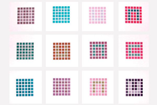
Each card is ruled in the form of a table and has an equal number of cells (6 pieces) vertically and horizontally. 10 cells differ from the rest in color and form a kind of square without one side. The task of the subject is to determine on which side of the square there is a gap.
The higher the card number, the greater the difference between the color of the text (a broken square or the letter "P") and the cells of the same tone that make up the background. The tables for deuteranopes and protanopes have 5, 10, 20, and 30 discrimination thresholds, respectively, as the number increases. Cards 9 through 11 for diagnosing tritanopia have 5, 10, and 15 discrimination thresholds.
The advantage of the threshold test is that it is impossible to falsify the results by learning the decoding of the images on the cards, which is widely practiced among those wishing to obtain a driving license, when the color perception test is carried out using Rabkin tables. People simply do not think about what consequences such falsification may lead to in the future.
But Yustova's tables also have one significant drawback. Print quality significantly affects the relevance of the results. Incorrect color rendering during printing led to some editions of Yustova's tables giving false results. Using inkjet printing would significantly reduce the number of deviations, but the price of the finished edition would then increase significantly, which would be unprofitable from the point of view of serial production.
For now, the market is dominated by cheap versions made using lithography, the quality control of which is highly questionable. Thus, a useful invention was effectively destroyed at the root.