Medical expert of the article

New publications
Electrooculography
Last reviewed: 07.07.2025

All iLive content is medically reviewed or fact checked to ensure as much factual accuracy as possible.
We have strict sourcing guidelines and only link to reputable media sites, academic research institutions and, whenever possible, medically peer reviewed studies. Note that the numbers in parentheses ([1], [2], etc.) are clickable links to these studies.
If you feel that any of our content is inaccurate, out-of-date, or otherwise questionable, please select it and press Ctrl + Enter.
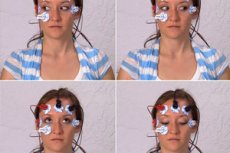
Electrooculography is the recording of the constant potential of the eye using skin electrodes placed on the area of the outer and inner edge of the lower eyelid. This method allows us to identify pathological changes in the pigment epithelium of the retina and photoreceptors. The method is based on the fact that the eye is a dipole: the cornea has a positive charge, the pigment epithelium is negative, and the existing constant potential changes when the eye moves under various adaptation conditions.
The study is conducted in a state of light and dark adaptation.
- Electrodes are placed on the skin at the medial and lateral edges.
- The patient is asked to rhythmically move his gaze from side to side with the same amplitude. With each movement of the eyeball, the electrode closest to the cornea becomes active relative to the other.
- The potential difference passes through the amplifier and is recorded.
The necessary conditions for normal light and dark oscillations of constant potential are normal functioning of photoreceptors and pigment epithelium, contact between these layers, and adequate blood supply to the choroid. The following indicators are noted in electrooculography:
- baseline potential - a constant potential measured in a patient who has been in constant illumination conditions for a long time;
- light rise potential: with a sharp change in light conditions from moderate illumination to bright light, a characteristic increase in the retinal base potential occurs (light rise);
- potential for a tempo decline: a sharp transition from moderate illumination to darkness leads to the emergence of a series of damped oscillations of the base potential (dark decline), reaching a minimum at the 10-12th minute of dark adaptation.
For clinical purposes, the ratio of the light peak potential to the dark decay potential is calculated. The result is multiplied by 100 to obtain the so-called Arden coefficient (AC), which is considered normal if it exceeds 185%. For the purpose of assessing pathological conditions of the retina, the AC is divided into subnormal (135-185%), abnormal (110-135%), extinguished (100-110%), and distorted (below 100%).
Electrooculography is used in the diagnosis of various retinal diseases of a dystrophic, inflammatory and toxic nature, in circulatory disorders and other pathologies in which photoreceptors and the choroid are involved in the pathological process.
 [
[ What do need to examine?
How to examine?