Medical expert of the article

New publications
Electroretinography
Last reviewed: 07.07.2025

All iLive content is medically reviewed or fact checked to ensure as much factual accuracy as possible.
We have strict sourcing guidelines and only link to reputable media sites, academic research institutions and, whenever possible, medically peer reviewed studies. Note that the numbers in parentheses ([1], [2], etc.) are clickable links to these studies.
If you feel that any of our content is inaccurate, out-of-date, or otherwise questionable, please select it and press Ctrl + Enter.
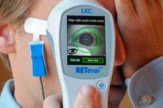
Electroretinography is a method of recording the total bioelectrical activity of all neurons in the retina: the negative a-wave of photoreceptors and the positive b-wave of hyper- and depolarizing bipolars and Müller cells. An electroretinogram (ERG) occurs when the retina is exposed to light stimuli of various sizes, shapes, wavelengths, intensities, durations, and repetition rates under various conditions of light and dark adaptation.
An electroretinogram records the retinal action potential in response to light stimulation of the appropriate intensity, i.e. the potential between the active corneal electrode embedded in a contact lens (or a film gold electrode fixed to the lower eyelid) and the reference electrode on the patient's forehead. An electroretinogram is recorded under conditions of light adaptation (photopic electroretinogram) and tempo adaptation (scotopic electroretinogram). Normally, an electroretinogram is biphasic.
- a-wave - the first negative deviation from the isoline, the source of which are photoreceptors.
- b-wave is a positive deflection generated by Müller cells and reflects the bioelectrical activity of bipolar cells. The amplitude of the b-wave is measured from the negative peak of the a-wave to the positive peak of the b-wave, increases with dark adaptation and with increasing brightness of the light stimulus; the b-wave consists of subcomponents: b1 (reflects the activity of rods and cones) and b2 (activity of cones). A special recording technique allows us to isolate rod and cone responses.
The practical value of electroretinography is determined by the fact that it is a very sensitive method for assessing the functional state of the retina, which allows one to determine both the most minor biochemical disorders and gross dystrophic and atrophic processes. Electroretinography helps to study the mechanisms of development of pathological processes in the retina, facilitates early differential and topical diagnostics of retinal diseases, it is used to monitor the dynamics of the pathological process and the effectiveness of treatment.
An electroretinogram can be recorded from the entire area of the retina and from a local area of varying size. A local electroretinogram recorded from the macular area allows one to evaluate the functions of the cone system of the macular area. An electroretinogram evoked by a reverse checkerboard stimulus is used to characterize a second-order neuron.
The allocation of functions of the photopic (cone) and scotopic (rod) systems is based on the difference in the physiological properties of the cones and rods of the retina, therefore, the corresponding conditions in which each of these systems dominates are used. Cones are more sensitive to bright red stimuli presented under photopic lighting conditions after preliminary light adaptation, suppressing rod activity, to a flicker frequency of over 20 Hz, rods - to weak achromatic or blue stimuli under dark adaptation conditions, to a flicker frequency of up to 20 Hz.
Varying degrees of involvement of the rod and/or cone systems of the retina in the pathological process are one of the characteristic signs of any retinal disease of hereditary, vascular, inflammatory, toxic, traumatic and other genesis, which determines the nature of the electrophysiological symptoms.
The classification of electroretinograms adopted in electroretinography is based on the amplitude characteristics of the main a- and b-waves of the electroretinogram, as well as their time parameters. The following types of electroretinograms are distinguished: normal, supernormal, subnormal (plus- and minus-negative), extinct, or unrecorded (absent). Each type of electroretinogram reflects the localization of the process, the stage of its development, and pathogenesis.
Normal electroretinogram
Includes 5 types of response. The first 3 types are recorded after 30 minutes of dark adaptation (scotopic), and 2 types - after 10 minutes of adaptation to diffuse illumination of average brightness (photopic).
Scotopic electroretinogram
- rod response to a low-intensity white flash or to a blue stimulus: high-amplitude b-wave and low-amplitude or undetectable a-wave;
- mixed rod and cone response to a high-intensity white flash: prominent a- and b-waves;
- oscillatory potentials to a bright flash and with special registration parameters. Oscillations are registered on the ascending "knee" of the b-wave and are generated by cells of the inner layers of the retina.
Photopic electroretinogram
- The cone response to a single bright flash consists of an a-wave and a b-wave with small oscillations;
- The cone response is used to record an isolated cone response when stimulated by a flickering stimulus with a frequency of 30 Hz, to which the rods are insensitive. The cone response is normally recorded for a flash of up to 50 Hz, above which individual responses are not recorded (critical flicker fusion frequency).
A supernormal electroretinogram is characterized by an increase in a- and b-waves, which is observed at the first signs of hypoxia, drug intoxications, sympathetic ophthalmia, etc. A supernormal bioelectrical reaction during a traumatic rupture of the optic nerve and its atrophy is caused by a violation of the conduction of excitation along the retino-thalamic centrifugal inhibitory fibers. In some cases, it is difficult to explain the nature of a supernormal electroretinogram.
Subnormal electroretinogram is the most frequently detected type of pathological electroretinogram, which is characterized by a decrease in a- and b-waves. It is recorded in dystrophic diseases of the retina and choroid, retinal detachment, uveitis with the involvement of the 1st and 2nd retinal neurons, chronic vascular insufficiency with impaired microcirculation, some forms of retinoschisis (X-chromosomal, sex-linked, Wagner syndrome), etc.
A negative electroretinogram is characterized by an increase or preservation of the a-wave and a small or significant decrease in the b-wave. A negative electroretinogram can be observed in pathological processes in which changes are localized in the distal parts of the retina. A minus-negative electroretinogram occurs in ischemic thrombosis of the central retinal vein, drug intoxications, progressive myopia and congenital stationary night blindness, Ogushi disease, X-chromosomal juvenile retinoschisis, retinal metalloses and other types of pathology.
An extinct or unrecorded (absent) electroretinogram is an electrophysiological symptom of severe irreversible changes in the retina with its total detachment, developed metallosis, inflammatory processes in the eye membranes, occlusion of the central retinal artery, and also a pathognomonic sign of pigment retinitis and Leber's amaurosis. The absence of an electroretinogram is noted with gross irreversible changes in neurons, which can be observed in dystrophic, vascular and traumatic lesions of the retina. An electroretinogram of this type is recorded in the terminal stage of diabetic retinopathy, when the gross proliferative process spreads to the distal parts of the retina, and in vitreoretinal dystrophy of Favre-Goldman and Wagner.
What do need to examine?
How to examine?
Who to contact?