Medical expert of the article

New publications
Ankle arthroscopy
Last reviewed: 04.07.2025

All iLive content is medically reviewed or fact checked to ensure as much factual accuracy as possible.
We have strict sourcing guidelines and only link to reputable media sites, academic research institutions and, whenever possible, medically peer reviewed studies. Note that the numbers in parentheses ([1], [2], etc.) are clickable links to these studies.
If you feel that any of our content is inaccurate, out-of-date, or otherwise questionable, please select it and press Ctrl + Enter.
According to domestic and foreign literature, ankle joint injuries account for 6 to 21% of musculoskeletal injuries. Despite the large arsenal of tools available to modern traumatologists, the high frequency of unsatisfactory treatment outcomes for this pathology with conservative treatment is 17%, with surgical treatment - 11%.
Damage to bone and soft tissue formations leads to the development of secondary changes in the joint, degenerative-dystrophic processes, structural reorganization of both damaged and intact tissues of the ankle joint, which ultimately leads to its functional insufficiency and contracture.
The radiographic picture of bone damage has been well studied. However, a number of intra-articular disorders cannot be determined using radiographic methods alone. These include ligament sprains, articular cartilage injuries in acute trauma, and in chronic trauma - chondromalacia, cysts, intra-articular bodies.
With open intervention, the risk of progression of joint pathology is aggravated: the occurrence of an inflammatory process, postoperative instability in the ankle joint, increasing limitation of movement, pain in the ankle, synovitis, contracture, and sometimes the development of ankylosis. Patients with various injuries of the ankle joint, as a rule, have a walking disorder, they experience pain when standing for a long time, and cannot wear regular shoes.
 [
[ Indications and contraindications for ankle arthroscopy
Indications for ankle arthroscopy are as follows:
- pain of unknown etiology;
- synovitis, hemarthrosis;
- joint blockades (intra-articular bodies);
- transchondral fractures and cartilage detachments;
- initial symptoms of deforming arthrosis;
- osteochondritis dissecans;
- cartilage changes in impingement syndrome;
- chondromatosis;
- arthritis;
- ankle fractures;
- joint instability;
- arthrodesis.
Relative contraindications:
- skin infection;
- inflammatory diseases in paraarticular tissues;
- severe stages of deforming arthrosis;
- complicated somatic condition of the patient.
Arthroscopic approaches
In diagnostic and operative arthroscopy of the ankle joint, three anterior and two posterior approaches are used, which are used in different combinations to introduce the arthroscope and instruments. The anterior approaches are located along the anterior joint space.
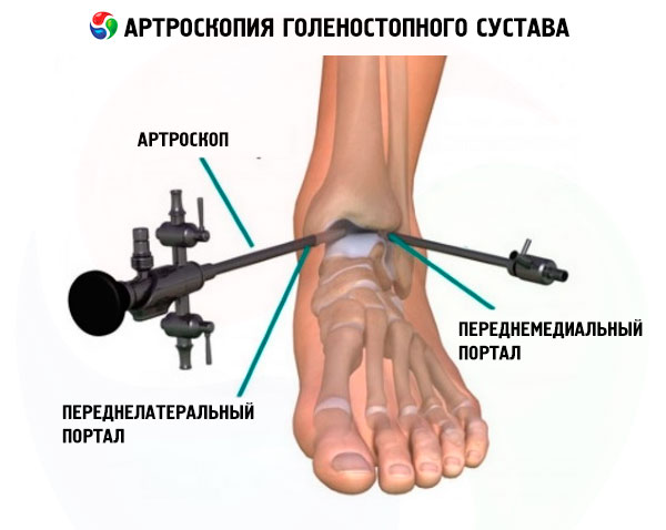
The anteromedial (anterior internal) approach is localized 0.5 cm below the joint space, somewhat medial to the tendon of the anterior tibialis muscle, lateral to the medial malleolus, proximal to the medial edge of the dome of the talus. There is a risk of damaging the terminal branch of the n. saphenous and v. saphenous.
The anterolateral (front-outer) approach serves as the main portal for performing arthroscopy. It is located 0.5 cm distal to the joint space, slightly lateral to the tendon of the fifth finger, medial to the lateral malleolus, proximal to the lateral part of the dome of the talus. Damage to the external cutaneous branch of the peroneal nerve is possible.
The anterocentral approach is located 0.5 cm distal to the joint space, between the long extensor of the great finger and the tendon of the anterior tibial muscle. There is a risk of damaging the deep peroneal nerve and the anterior tibial artery.
The posterolateral (posterolateral) approach is the only recommended posterior portal. It is located 1 cm below the anterior approaches and 0.5 cm distal to the joint space, adjacent to the Achilles tendon. Damage to the v. saphenous and n. surahs is possible.
The posteromedial (rear-internal) approach is located 0.5 cm distal to the joint space, slightly medial to the edge of the Achilles tendon at this level. This approach is not recommended due to its ineffectiveness and high risk of damaging the tarsal canal structures (posterior tibial nerve and artery).
A fairly complete view of the ankle joint is possible from two anterolateral approaches using a 4.5 mm diameter arthroscope with a 30° viewing angle.
Using the listed approaches, it is possible to examine 95% of the joint space: the articular surfaces of the tibia and talus, both ankles, talomaxillary joints, deltoid ligament, talofibular ligaments, synovial pockets.
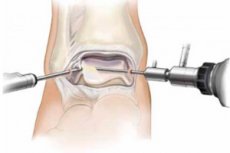
Technique for performing ankle arthroscopy
The procedure is performed under spinal or conduction anesthesia. The patient's position on the operating table is supine. The limb to be operated on is fixed at the level of the middle third of the shin and secured to the operating table in a special support at a height of 20 cm. After the surgical field has been treated, arthroscopy of the ankle joint is performed from two approaches: anteromedial and anterolateral. At the same time, the assistant stretches the joint space of the ankle joint by traction on the foot (manual distraction method). Other distraction methods can also be used: distraction by cuff traction (using a weight) and with the help of devices and accessories (for example, a rod distractor). The optimal distraction value is 7-8 mm.
First, the anterior and then the posterior part of the joint are examined. After the arthroscope is inserted into the ankle joint cavity, the articular surfaces of the tibia and talus, both malleoli, talomaxillary joints, deltoid ligament, talofibular ligaments, and synovial pockets are examined. In the case of initial signs of deforming arthrosis, high-frequency ablation and shaving of the articular surface are performed; if intra-articular bodies are present, they are removed. In case of dissecting osteochondritis of the talus, high-frequency ablation of the cartilage of the talus is used.