Medical expert of the article

New publications
Diagnosis of osteoarthritis: arthroscopy
Last reviewed: 06.07.2025

All iLive content is medically reviewed or fact checked to ensure as much factual accuracy as possible.
We have strict sourcing guidelines and only link to reputable media sites, academic research institutions and, whenever possible, medically peer reviewed studies. Note that the numbers in parentheses ([1], [2], etc.) are clickable links to these studies.
If you feel that any of our content is inaccurate, out-of-date, or otherwise questionable, please select it and press Ctrl + Enter.
Today, osteoarthritis treatment is mainly aimed at improving symptoms, primarily pain relief. Current research is developing drugs that can change the course of osteoarthritis: prevent, delay the development of changes in the joints, or even cause their regression. Such research requires standardized and reproducible assessments of changes in the joints to clearly assess the results of treatment. This primarily concerns the assessment of the quantity, integrity, and/or quality of articular cartilage.
In recent years, arthroscopy has been considered as a method for early diagnostics of osteoarthrosis, since it allows for the detection of the above-mentioned cartilage changes even in the absence of radiographic signs of the disease. For example, when applied to the knee joint, this method provides direct, including magnification, visualization of the six surfaces of the joint, and the technique is more sensitive than radiography or MRI in relation to cartilage damage. The advantages of arthroscopy have led to this method being considered the "gold standard" for assessing the condition of articular cartilage. Some authors, taking these advantages into account, call the technique "chondroscopy". Direct visualization allows for the assessment of the synovial membrane, the severity of synovitis, and also for targeted biopsy, which is of particular importance for the anterior parts of the knee joint, where changes in osteoarthrosis are often fragmentary.
The main problems of arthroscopy today include the following: its invasive nature, insufficiently developed standardized assessment systems for chondropathy in osteoarthritis, as well as recommendations for the unification of visualization of articular cartilage surfaces.
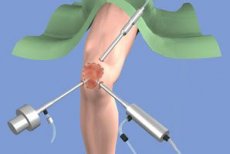
Arthroscopy technique
Arthroscopy performed for therapeutic purposes is often performed under general or spinal anesthesia, while diagnostic arthroscopy can be performed under local (subcutaneous or intra-articular) anesthesia, which makes the procedure safer, more accessible and inexpensive. E. Eriksson et al. (1986), when comparing the results of various arthroscopy techniques, found that about 77% of patients were satisfied with the procedure under local or spinal anesthesia, while 97% were satisfied with the procedure under general anesthesia. PM Blackburn et al. (1994) found good tolerability of arthroscopy performed under local anesthesia, comparable to MRI of the knee joints, in all 16 patients examined, with 8 of them preferring arthroscopy, 2 - MRI, and 6 reporting equally good tolerability of both procedures.
In a prospective study by X. Ayral et al. (1993), 84 patients underwent chondroscopy under local anesthesia, and tolerance was rated as "good" by 62% of patients, "very good" by 28%. 25% of these patients did not feel any pain at all, and 75% noted minor pain during the procedure or immediately after it. Daily motor activity after arthroscopy was difficult in 79% of patients (up to 1 day - in 44%, up to 2 days - in 55%, up to 1 week - in 79%). By the end of the 1st month after chondroscopy, 82% of patients noted an improvement in their condition.
JB McGintyn RA Matza (1978) evaluated the diagnostic accuracy of arthroscopy performed under general or local anesthesia using post-arthroscopic visualization through arthrotomy. It was found that arthroscopy was slightly more accurate when performed under local anesthesia (95%) than under general anesthesia (91%). However, it should be emphasized that performing arthroscopy under local anesthesia requires more training, even for experienced arthroscopists.
Arthroscope with small glass lens
Knee arthroscopy is often performed using an arthroscope with a 4 mm glass lens and a 5.5 mm trocar. In some patients with ligament contractures or residual muscle tightness (due to local anesthesia), the posterior tibiofemoral joint may be inaccessible to a standard arthroscope (4 mm). An arthroscope with a 2.7 mm lens has a field of view comparable to a standard arthroscope and allows examination of all joint compartments in most cases. Continuous irrigation of the knee joint provided by a 2.7 mm arthroscope is sufficient to clear the joint of blood and various particles and provide a clear field of view for visualization. Technically, a 25-30° field of view provides a wide and better view. Smaller diameter fiberoptic arthroscopes (1.8 mm) can be inserted into the joint through a puncture hole rather than an incision, but they have several disadvantages: a smaller field of view, a dimmer and grainier image due to image transfer along the fibers and poorer irrigation, and a tendency for the optical fibers to bend and break, often resulting in only a direct image. According to these authors, the sensitivity of such arthroscopes compared with standard ones in detecting cartilage defects is 89%, and for synovial membrane defects - 71%.
The results of a prospective, open, uncontrolled study by X. Ayral et al. (1993) indicate an improvement in well-being in 82% of patients 1 month after chondroscopy. It is believed that lavage of the joint cavity performed during the procedure (usually about 1 liter of isotonic sodium chloride solution) provides clinical improvement in the manifestations of the joint syndrome, which is confirmed by the data of controlled studies, and eliminates the potential harm of this invasive procedure.
 [ 7 ]
[ 7 ]
Arthroscopic assessment of the severity of cartilage damage in osteoarthritis
[ 8 ], [ 9 ], [ 10 ], [ 11 ], [ 12 ]
Traditional classification systems
To assess the dynamics of articular cartilage damage in osteoarthrosis, especially under the influence of treatment, quantitative assessment systems are needed that provide three main parameters of these lesions: depth, size and localization. Many different arthroscopic classification systems are currently known.
Some classification systems consider only the depth of articular cartilage lesions and provide qualitative information about the cartilage surface without providing a quantitative approach to the recording of cartilage lesions. Other systems consider a combination of the depth and the size of the most severe articular surface chondropathy in a single descriptive category, but there are many discrepancies. A brief description of the classification systems is given below.
The classification system proposed by R. E. Outerbridge (1961) divides cartilage damage into degrees:
- Grade I - softening and swelling of the cartilage without cracks (true chondromalacia);
- II - fragmentation of cartilage and formation of cracks with a diameter of 0.5 inches or less;
- III - fragmentation of cartilage and formation of cracks with a diameter of more than 0.5 inches;
- IV - cartilage erosion involving subchondral bone.
It is evident that grades II and III have the same depth and the size is described for them, while grades I and IV are not assessed in detail. In addition, the size of cracks (grades II and III) is not a constant value.
R. P. Ficat et al. (1979) divided cartilage lesions into closed and open chondromalacia, with closed chondromalacia (grade I) representing true chondromalacia (softening and swelling) and open chondromalacia (grade II) representing open (with fissures) chondropathy. According to this system, a grade I lesion starts with a 1 cm2 area and progressively extends in all directions. This description leads to inconsistency in the question of the total area of cartilage surface area affected. Grade II includes three different depths of chondropathy: superficial and deep fissures and involvement of the subchondral bone without specifying the dimensions. Consequently, this system lacks a precise quantitative approach to assessing the degree of articular cartilage destruction.
Characteristics of classification systems for arthroscopic assessment of articular cartilage lesions
Author |
Description of the articular cartilage surface |
Diameter |
Localization |
RE Outer Ridge, 1961 |
I - thickening and swelling |
I - no description |
Begins most often on the medial surface of the patella; then "mirror-like" spreads to the lateral surface of the intercondylar region of the femoral condyles; upper edge of the medial condyle of the femur |
II - fragmentation and crack formation |
II - less than 0.5 inches |
||
III - fragmentation and crack formation |
III - more than 0.5 inches |
||
IV - erosion of cartilage and subchondral bone |
IV - no description |
||
SW Cassels, 1978 |
I - superficial erosions of cartilage |
I-1 cm and less |
Patella and anterior surfaces of the femur |
II - deeper erosions of cartilage |
II -1-2 cm |
||
III - the cartilage is completely eroded, subchondral bone is involved |
III - 2-4 cm |
||
IV - articular cartilage is completely destroyed |
IV - "wide area" |
||
RP Float et al. 1979 |
I - closed chondromalacia; simple thickening (simple bullae) macroscopically, surface intact, varying degrees of severity from simple thickening to "deep edema", loss of elasticity |
I - 1 cm, then the lesion spreads progressively in all directions |
Lateral surface |
II - open chondromalacia: A) cracks - single or multiple, relatively shallow or extending to the subchondral bone B) Ulceration - localized "loss" of cartilaginous substance involving subchondral bone. The bone surface may appear "polished" (eburnation of bone) |
II - no description |
Medial surface (violation of articular relationships of 2° or more) |
|
Formation of cartilage "fragments" - multiple, separated from each other by deep cracks extending to the subchondral bone. Superficial changes - fraying of cartilage; longitudinal grooves defined along the axis of joint movement. |
Not localized, but the entire contact area is involved |
Centered on the ridge separating the medial and distal surfaces |
|
J. Beguin, B. Locker, 1983 |
I - softening, swelling II - surface cracks III - deep cracks extending to the subchondral bone IV - subchondral bone involvement |
Description is missing |
Description is missing |
JNInsall, 1984 |
I - swelling and softening of the cartilage (closed chondromalacia) II - deep cracks extending to the subchondral bone III - delamination IV - erosive changes and involvement of the subchondral bone (osteoarthritis) |
Description is missing |
I-IV: center of the patella crest with equal extension to the medial and lateral surfaces of the patella IV: opposite or "mirror" surfaces of the femur are also involved. The upper and lower thirds of the patella are often slightly damaged, the femur is slightly involved |
I - fraying or cracking |
I - less than 0.5 cm |
Most often at the junction of the medial and distal surfaces of the patella |
|
II - fraying or cracking |
II - 0.5-1 cm |
||
III - fraying or cracking |
III -1-2 cm |
||
IV - delamination with or without subchondral bone involvement |
IV - more than 2 cm |
In the classification proposed by G. Bently, J. Dowd (1984), degrees I, II and III have the same features (fibrilization or crack formation), and the differences between the degrees are based on the diameter of the lesions. There is no mention of true chondromalacia. Degree IV corresponds to two different depths of chondromalacia: fibrilization with or without involvement of the subchondral bone, with a fixed size of more than 2 cm. A reasonable question arises, what degree of lesion corresponds to involvement of the subchondral bone with a diameter of less than 2 cm?
SW Cassels (1978) assessed the diameter of lesions in centimeters and the relative depth of lesions, initially assuming that a smaller lesion depth corresponds to a smaller diameter. In this case, what degree corresponds to superficial lesions involving the entire articular surface?
Thus, the above systems do not provide sufficient information about the depth, size, and location of cartilage damage. In addition, the scoring system must be applicable to both the knee joint as a whole and to each of its three compartments: patellofemoral, medial, and lateral tibiofemoral. However, without quantitative joint mapping, the description of the location of chondropathy outside a given articular surface remains qualitative.
Modern classification systems
In 1989, FR Noyes and CL Stabler proposed their own grading system for articular cartilage damage. They divided the description of the articular surface (cartilage/subchondral bone), the depth of the lesion, the diameter and localization of the lesion. The authors distinguish three degrees of articular surface damage: Grade 1 - the articular surface is intact; Grade 2 - the articular surface is damaged, open lesion; Grade 3 - bone involvement. Each of these degrees is divided into types A or B depending on the depth of the lesion. Grade 1 implies chondromalacia. Type 1A corresponds to a moderate degree of softening of the articular cartilage; type 1B - significant softening with swelling of the articular surface. Grade 2 is characterized by any destruction of the articular surface without visualized bone involvement. Type 2A lesions include superficial cracks (less than half the thickness of the cartilage); Type 2B - more than half the thickness (deep cracks down to the bone). Grade 3 indicates bone involvement. Type 3A suggests that the normal bone contour is preserved; type 3B - indicates cavitation or erosion of the bone surface. All detected lesions are marked on the knee joint diagram, and the diameter of each is estimated by the examiner in millimeters using a special graduated "hook". Depending on the diameter and depth of the lesion, a point scale is used to quantify the severity of chondropathy for each joint section and ultimately to perform a total joint count.
The FR Noyes, CL Stabler system was the first attempt by researchers to quantify chondropathy, so it is not without its drawbacks:
- All cartilage lesions are represented on knee diagrams as a complete circle with the diameter determined by a graduated "hook". This is not a very objective method of estimating size, since most cartilage lesions are not strictly circular, but are often oval or have no definite shape. In addition, degenerative changes in cartilage can often have a shape with the deepest lesion in the center, surrounded by a zone of more superficial cartilage damage; and to this "surrounding lesion", which has a crown shape, the diameter cannot be applied.
- Any lesion smaller than 10 mm in diameter is not considered clinically significant, which leads to a loss of sensitivity of the technique. When monitoring the effect of the basic drug, any, even the smallest, lesions should be described.
- The point scale for assessing both the depth and diameter of cartilage lesions is arbitrary and is not based on statistical methodology or clinical assessment and consideration of the severity of these lesions.
The newest of the proposed methods of arthroscopic assessment of chondropathy were proposed by H. Auga1 and co-authors (1993, 1994), M. Dougados and co-authors (1994).
The first of these methods is based on the subjective global assessment of chondropathy by the examiner; it is based on a 100-mm visual analogue scale (VAS), with "0" representing no chondropathy and "100" representing the most severe chondropathy. One VAS is used for each articular surface of the knee: the patella, trochlea, medial and lateral condyles, and medial and lateral tibial plateau. A VAS score is obtained for each of the three knee compartments and is obtained by averaging the VAS scores for the two corresponding articular surfaces of the joint compartment.
The second method is more objective and is based on an analytical approach, which includes a joint diagram of the knee joint with gradation of the localization, depth and size of all existing cartilage damage.
Localization
The technique includes 6 zones of determination: patella, block (intercondylar fossa), medial and lateral condyles (separately), medial and lateral plateau of the tibia (separately).
Depth
The system is based on the classification of chondropathy proposed by French arthroscopists J. Beguin, B. Locker (1983), in which 4 degrees of cartilage damage are distinguished:
- Grade 0 - normal cartilage;
- Grade I - chondromalacia including softening with or without edema; may correspond to grade 1, types A and B according to FR Noyes, CL Stabler (1989);
- Grade II - the cartilage contains superficial cracks, single or multiple, giving the surface a "velvety" appearance; this grade also includes superficial erosions. The cracks and erosions do not reach the surface of the subchondral bone. May correspond to grade 2Apo FR Noyes, CL Stabler, 1989 (i.e., lesions occupying less than half the thickness of the cartilage);
- Grade III - there are deep cracks in the cartilaginous surface down to the subchondral bone that are not directly visualized but can be identified with an arthroscopic probe; Grade III may be in the form of a "shark's mouth" or a separate piece of cartilage formed due to a single deep crack, "crab meat" due to multiple deep tears. Grade III also includes deep ulceration of the cartilage, forming a crater that remains covered by a thin layer of cartilage. May correspond to grade 2B according to FR Noyes, CL Stabler, 1989 (i.e. lesions occupying more than half the thickness of the cartilage);
In osteoarthritis of the knee joint, the destruction of articular cartilage often manifests itself as a combination of varying degrees of severity, with the most severe areas of damage surrounded by areas of less severe damage.
To create a unified chondropathies score, a multivariate analysis was used using logistic multiple regression, in which the dependent variable was the overall assessment of chondropathies by the investigator using VAS, and the independent variables were the depth and size of the lesions. Thus, two chondropathies scoring systems were created: the SFA scoring system and the SFA grading system.
SFA score is a variable with values from “0” to “100”, obtained for each joint section using the formula:
SFA score = A + B + C + D,
Where A = size (%) of first degree damage x 0.14;
B = size (%) of grade II damage x 0.34;
C = size (%) of grade III damage x 0.65;
D = size (%) of grade IV damage x 1.00.
Size (%) = average percentage of surface area of the medial femoral condyle and medial tibial plateau (medial tibiofemoral compartment - TFC), lateral femoral condyle and lateral tibial plateau (lateral TFC), or trochlea and patella (patellofemoral compartment - PFC).
The severity coefficients of chondropathy (0.14; 0.34; 0.65; 1.00) were obtained by parametric multivariate analysis.
SFA grade is a semi-quantitative value. The above values (size (%) of grades I-IV lesions) are substituted into the formula to obtain the total grade (or severity category of chondropathy of the department) for each of the knee joint departments. The formula for each department was obtained by nonparametric multivariate analysis using regression analysis; in total - 6 categories for the PFO (0-V) and 5 categories for the medial and lateral TFO (0-IV). An example of calculating the SFA score and SFA grade is presented in Table 20.
[ 13 ], [ 14 ], [ 15 ], [ 16 ], [ 17 ]
ACR system
In 1995, the ACR committee proposed a scoring system for cartilage. This system takes into account the depth, size, and location of cartilage damage and then enters the data into a knee diagram. The depth of each damage is graded (Noyes FR, Stabler CL, 1989 classification); the size of each damage is expressed as a percentage. A point scale is used to calculate the overall score, the so-called damage score. The reliability of the latter was assessed by D. Klashman et al. (1995) in a blinded study: videotapes of 10 arthroscopies were viewed twice by three rheumatologists-arthroscopists, and high reliability was demonstrated both for the data of one expert in two studies (r = 0.90; 0.90; 0.80; p < 0.01 for each) and between experts (r = 0.82; 0.80; 0.70; p < 0.05 for each).
[ 18 ], [ 19 ], [ 20 ], [ 21 ]
Comparative analysis of reliability, significance and sensitivity to changes of arthroscopic SFA, VAS systems
X. Ayral et al. (1996) found a close correlation between arthroscopic quantitative assessment of chondropathy and radiographic assessment of joint space narrowing under weight-bearing conditions, namely the following indicators:
- overall assessment of chondropathy (VAS) and narrowing of the radiographic joint space (RSS) of the medial part of the joint, expressed as % (r = 0.646; p < 0.0001);
- SFA score and narrowing of the SRSF in the medial and lateral TFO, expressed in mm (r = -0.59; p<0.01 and r = -0.39; p<0.01, respectively);
- SFA grade and medial and lateral TFO RSM narrowing expressed in mm (r = -0.48; p < 0.01 and r = -0.31; p < 0.01, respectively). Despite these results, arthroscopy was more sensitive than radiography: even deep and extensive cartilage erosions may remain undetected on radiographs, even with weight-bearing radiography. Of 33 patients with ACR-conclusive osteoarthritis who had medial TFO RSM narrowing < 25% on weight-bearing radiography, 30 had chondropathy on arthroscopy with a mean VAS score of 21 mm (2–82 mm), including > 10 mm in 24 patients.
X. Ayral et al. (1996) found a statistically significant correlation (p<0.05) between articular cartilage damage: 1) of three sections of the knee joint (medial, lateral, PFO) and the age of patients; and 2) of the medial section of the joint and body mass index. When conducting repeat arthroscopy after 1 year (41 patients), the same authors showed that changes in the severity of cartilage damage correlated with changes in functional insufficiency of the musculoskeletal system (Lequesne index: r = 0.34; p = 0.03) and quality of life (AIMS2: r = 0.35; p = 0.04). In the same study, the VAS score of the medial joint changed from 45±28 at the beginning of the study to 55+31 after 1 year (p = 0.0002), and the SFA score - from 31+21 to 37+24 (p = 0.0003). Similar results, indicating high sensitivity of arthroscopy to dynamic changes in cartilage, were also obtained by Y. Fujisawa et al. (1979), T. Raatikainen et al. (1990), and V. Listrat et al. (1997) during dynamic arthroscopic assessment of changes in the articular cartilage of patients with osteoarthritis under the influence of chondroprotective therapy (hyaluronan).
The use of microscopic technology allows arthroscopic visualization of changes that are inaccessible to other research methods.
Thus, arthroscopy performed under local anesthesia is an adequate method for instrumental diagnosis of osteoarthritis and can also be used to monitor the effectiveness of treatment, primarily with disease-modifying drugs (DMOAD).