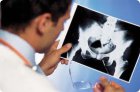
Ensuring effective therapy for osteoarthritis and disease relapses largely depends on the use of standardized approaches to its diagnosis and differential diagnosis.
Recent advances in specific and sensitive biochemical markers reflecting the overall rate of bone formation and resorption have significantly improved noninvasive assessment of bone metabolism in various metabolic bone diseases. Biochemical markers are generally classified as those for bone formation and those for bone resorption.
Significant advances in understanding the pathophysiology and evolution of osteoarthritis have led not only to improved diagnosis but also to a reassessment of the methodology and metrology of clinical trials in osteoarthritis. Clinical diagnosis of osteoarthritis is challenging.
In most cases, patients with osteoarthritis do not have changes in blood and urine tests, with the exception of cases of synovitis with significant effusion, when an increase in ESR, hypergammaglobulinemia, and an increase in the level of acute phase indicators - CRP, fibrinogen, etc. may occur.
The shoulder joint is one of the most convenient for ultrasound, especially because many pathological changes occur in its soft tissues. Due to the limited information available from radiographic imaging in reflecting soft tissue changes, ultrasound, along with MRI, has become the leading method for examining the shoulder joint.
Although MRI is the leading method for detecting coxarthrosis, ultrasound has advantages in detecting small effusions in the hip joint (even less than 1 ml), as well as disorders of the periarticular soft tissues in the early stages of osteoarthritis.
As is well known, radiography in most cases can detect knee joint damage when bone elements are involved in the pathological process. These changes are often irreversible, making treatment of such patients difficult.
Radioisotope joint scintigraphy is performed using osteotropic radiopharmaceuticals (pyrophosphate, phosphonium, labeled with 99mTc). These drugs actively accumulate in areas of active bone and collagen metabolism.
The use of ultrasound (sonography) in rheumatology is a relatively new and promising area. Over the past decade, ultrasound has become widely used as a visualization technique for examining patients with rheumatic joint diseases and for treatment monitoring.
MRI provides objective and quantitative measurements of subtle, undetectable morphological and structural changes in various joint tissues over time and is therefore a more reliable and easily reproducible method that helps monitor the progression of osteoarthritis.