Medical expert of the article

New publications
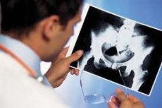
Hip ultrasound for osteoarthritis
Last reviewed: 08.07.2025

All iLive content is medically reviewed or fact checked to ensure as much factual accuracy as possible.
We have strict sourcing guidelines and only link to reputable media sites, academic research institutions and, whenever possible, medically peer reviewed studies. Note that the numbers in parentheses ([1], [2], etc.) are clickable links to these studies.
If you feel that any of our content is inaccurate, out-of-date, or otherwise questionable, please select it and press Ctrl + Enter.
Although the leading method for detecting coxarthrosis is MRI, ultrasound has advantages in detecting small effusions in the hip joint (even less than 1 ml), as well as disorders of the periarticular soft tissues in the early stages of osteoarthritis. The study is carried out using a linear or convex sensor in the range of 3.5-7 MHz, depending on the constitutional features of the patient.
The examination is usually performed from the anterior approach (longitudinal and transverse positions of the sensor), with the patient lying on his back with straight legs. The bone landmarks are the upper edge of the acetabulum and the semicircle of the femoral head. The hypoechoic hyaline cartilage and synovial joint capsule of the hip joint (represented by fibers of the ischiofemoral, pubofemoral and iliofemoral ligaments) are well visualized from the anterior approach. The lateral approach is used to visualize the greater trochanter and the trochanteric bursa, located superficially subcutaneously above it. The ischial tuberosity is examined from the posterior approach with the patient lying on his side with the limb being examined bent and brought to the stomach.
In one study, ultrasound was performed on 54 patients with osteoarthritis of the hip joints (diagnostic criteria AC R, 1990) aged 41 to 74 years (mean age 56.44±7.12 years); of which 22 were men and 32 were women; the duration of the disease was 0.6 years to 37 years (on average 8.3±3.48 years).
The presence of effusion in the hip joint was diagnosed if the distance between the surface of the femoral neck and the joint capsule exceeded 9-10 mm.