Medical expert of the article
New publications
Ultrasound of knee joints in osteoarthritis
Last reviewed: 08.07.2025

All iLive content is medically reviewed or fact checked to ensure as much factual accuracy as possible.
We have strict sourcing guidelines and only link to reputable media sites, academic research institutions and, whenever possible, medically peer reviewed studies. Note that the numbers in parentheses ([1], [2], etc.) are clickable links to these studies.
If you feel that any of our content is inaccurate, out-of-date, or otherwise questionable, please select it and press Ctrl + Enter.

As is known, radiography in most cases allows to determine the damage of the knee joint when bone elements are involved in the pathological process. Often these changes are already irreversible, treatment of such patients is difficult.
The advantages of ultrasound of the knee joint are accessibility, cost-effectiveness, absence of radiation exposure to the patient, the ability to visualize soft tissue components of the joint, allowing to identify early signs of lesions that are practically not determined by radiography.
The ultrasound technique developed by L. Rubaltelly (1993) allows determining the main signs of knee joint pathology - traumatic injuries, degenerative-dystrophic and inflammatory processes, etc.
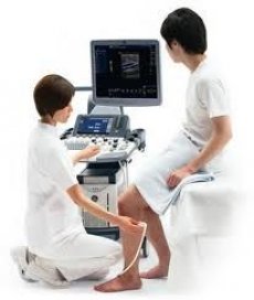
Ultrasound usually begins with the suprapatellar region. Here, the tendon of the quadriceps femoris, the contours of the upper pole of the patella, and the suprapatellar bursa (upper fold) are well visualized with longitudinal and transverse scanning. The study of this bursa in osteoarthrosis is especially informative for diagnosing the severity of degenerative-dystrophic and inflammatory lesions. Normally, the synovial membrane is not visualized. In deforming osteoarthrosis with synovitis, an increase in the bursa, straightening of folds, and the presence of excess fluid are noted.
Further examination with knee flexion and transverse position of the sensor allows visualization of the PFO of the joint, in particular the hyaline cartilage and the presence or absence of excess fluid above it. Moving the sensor to the area below the patella makes it possible to determine the superficially located patellar ligament, its structure, the infrapatellar fat pad, the infrapatellar synovial fold, deeper than which the anterior cruciate ligament is located. The transverse position of the sensor allows visualization of the articular cartilage of the lateral and medial condyles, changes in the shape of the articular surfaces of the femur (flattening, etc.). Placing the sensor on the inner and outer lateral surfaces of the knee joint allows visualization of the inner and outer collateral ligaments, marginal bone growths of the femur and tibia, the presence or absence of effusion, respectively.
With ultrasound of the popliteal fossa, it is possible to visualize pathological formations in this area (Baker's cyst), articular cartilages of the lateral and medial condyles, posterior parts of the medial and lateral condyles, posterior horns of the lateral and medial menisci, and the posterior cruciate ligament.
In one of the studies, 62 patients with gonarthrosis were examined, and a comparative assessment of ultrasound and thermography data was performed. Ultrasound of the musculoskeletal system was performed on a SONOLINE Omnia (Siemens) device with a 7.5L70 linear sensor (frequency 7.5 MHz) in the "ortho" mode in standard positions. The condition of the articular bone surfaces (including the condition of the cortical layer, including the subchondral bone), joint spaces, periarticular soft tissues, the presence of effusion and its characteristics, changes in the ligament-tendon apparatus and some other parameters were assessed.
According to ultrasound data, patients with osteoarthritis of the knee joints had: narrowing of the joint space due to a decrease in the height of the articular cartilage (transverse position of the sensor), bone growths (osteophytes) and/or defects of the articular surfaces of the bones, changes in the synovial membrane and the presence of effusion in the joints, changes in the paraarticular soft tissues (all positions). Changes in the surface of the cortical layer of the articular surfaces (unevenness, formation of surface defects) were recorded already at the initial stages of the disease (I radiographic stage according to Kellgren) and reached their maximum expression at stages III and IV.
Joint effusion was observed in 28 (45.16%) patients with gonarthrosis, mainly in stages II and III of the disease, it was mainly localized in the superior recess (in 32.3% of patients), in the lateral part of the joint space (in 17.7%), less often in the medial part of the joint space (in 9.7%) and in the posterior recess (in 3.2%)
The effusion had a homogeneous anechoic echostructure provided that the clinical symptoms of osteoarthrosis lasted up to 1 month, and in patients with clinical signs of persistent inflammation in the joint - non-homogeneous, with inclusions of various sizes and echodensities. The thickness of the synovial membrane was increased in 24 (38.7%) examined patients, and its uneven thickening was recorded in 14 of them. It should be noted that the average duration of the disease in these patients was longer than in the group of patients with gonarthrosis as a whole (6.7 + 2.4 years), and in patients with uneven thickening of the synovial membrane it was even longer (7.1 + 1.9 years). Thus, the features of synovitis reflected the duration of gonarthrosis and the severity of the process at the time of examination.
The assessment of the hyaline cartilage of the joint (subpatellar approach, transverse position of the sensor) was performed according to the following criteria: thickness, uniformity of thickness, structure, surface, changes in the surface of the subchondral bone (presence of cysts, erosions, other defects). The height of the cartilage decreased more on the medial condyle in accordance with the greater mechanical load on this area.
The results obtained by comparing data from remote thermography and ultrasound are noteworthy.
A strong or very strong direct relationship according to the correlation analysis data was found between the temperature gradient in the medial and lateral areas of the knee joints, on the one hand, and joint effusion and synovial membrane thickening according to ultrasound data, on the other. A weaker relationship was found between the presence of bone growths in the medial area of the knee joints (ultrasound data) and the temperature gradient in all examined areas of the joints.
Therefore, ultrasound and thermography are complementary methods in the diagnosis of osteoarthritis of the knee joints, which especially concerns the activity of the process and the severity of degenerative changes in the joints.

