Medical expert of the article

New publications
Tibia
Last reviewed: 06.07.2025

All iLive content is medically reviewed or fact checked to ensure as much factual accuracy as possible.
We have strict sourcing guidelines and only link to reputable media sites, academic research institutions and, whenever possible, medically peer reviewed studies. Note that the numbers in parentheses ([1], [2], etc.) are clickable links to these studies.
If you feel that any of our content is inaccurate, out-of-date, or otherwise questionable, please select it and press Ctrl + Enter.
The tibia is the thickest bone of the leg. The proximal end of the bone is thickened and forms the medial and lateral condyles (condylus medialis et condylus lateralis). The superior articular surface (facies articularis superior) faces upward and articulates with the condyles of the femur. Between the articular surfaces of the condyles of the tibia is the intercondylar eminence (eminentia intercondilaris), which consists of two tubercles: the medial intercondylar tubercle (tuberculum intercondylare mediale) and the lateral intercondylar tubercle (tuberculum intercondylare laterale). In front of the intercondylar eminence is the anterior intercondylar field (drea intercondyldris anterior), behind - the posterior intercondylar field (area intercondylaris). Below the lateral condyle, on its lateral side and slightly to the rear, there is a fibular articular surface (facies articularis fibularis) for connection with the fibula.
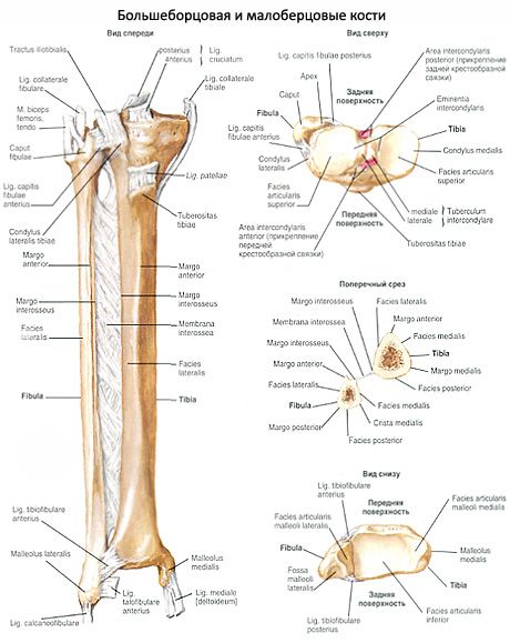
The body of the tibia (corpus tibiae) has a sharp anterior edge (margo anterior), which can be felt through the skin. The anterior edge thickens at the top and forms the tibial tuberosity (tuberositas tibiae), to which the quadriceps femoris is attached. The lateral edge is also sharp and faces the fibula. Therefore, it is called the interosseous edge (margo interosseus). The medial edge (margo medialis) is rounded. The body of the tibia has three surfaces. The medial surface is smooth and lies directly under the skin. The lateral and posterior surfaces are covered with muscles. On the posterior surface, a rough line of the soleus muscle (linea musculi solei) is visible, which runs from the posterior edge of the lateral condyle obliquely downwards and medially.
The lower distal end of the tibia is widened. On the lateral edge of the distal end of the bone is the fibular notch (incisura fibularis) for articulation with the fibula. On the medial side, the medial malleolus (malleolus medialis) extends downward. Behind it is a shallow malleolar groove (sulcus malleolaris) for the tendon of the posterior tibial muscle passing here. On the lateral side of the medial malleolus is the articular surface (facies articularis malleoli), which passes at an angle into the lower articular surface (facies articularis inferior) of the tibia. These surfaces, together with the articular surface of the fibula, articulate with the talus of the tarsus (foot).
What do need to examine?
How to examine?