Medical expert of the article
New publications
Tibia bones
Last reviewed: 06.07.2025

All iLive content is medically reviewed or fact checked to ensure as much factual accuracy as possible.
We have strict sourcing guidelines and only link to reputable media sites, academic research institutions and, whenever possible, medically peer reviewed studies. Note that the numbers in parentheses ([1], [2], etc.) are clickable links to these studies.
If you feel that any of our content is inaccurate, out-of-date, or otherwise questionable, please select it and press Ctrl + Enter.
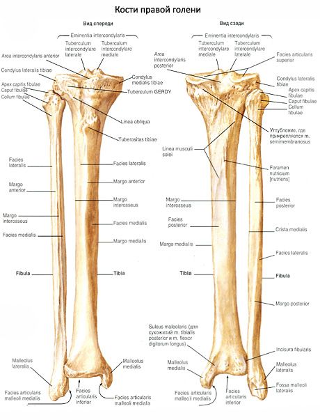
Bones of the lower leg. The lower leg has two bones. The tibia is located medially, and the fibula is located laterally. Each bone has a body and two ends. The ends of the bones are thickened and have surfaces for articulation with the femur at the top (tibia) and with the bones of the foot at the bottom. Between the bones is the interosseous space of the lower leg (spatium interosseum cruris).

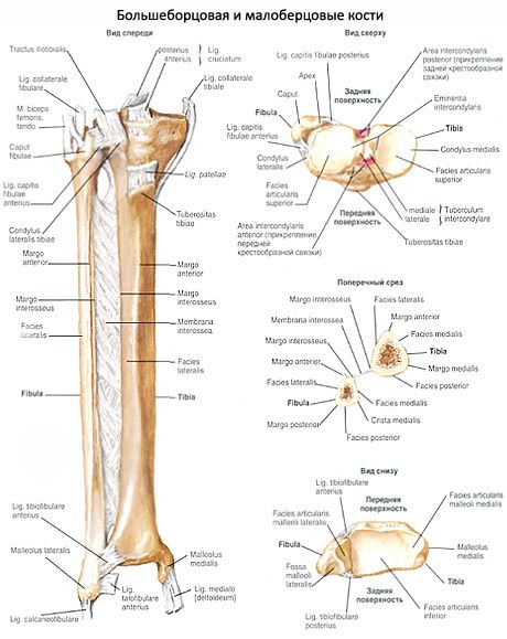
The bones of the leg are connected by the tibiofibular joint, as well as by continuous fibrous connections - the tibiofibular syndesmosis and the interosseous membrane of the leg.
The tibiofibular joint (art. tibiofibularis) is formed by the articulation of the articular fibular surface of the tibia and the articular surface of the head of the fibula. The articular surfaces are flat. The joint capsule is tightly stretched, reinforced in front by the anterior and posterior ligaments of the head of the fibula (ligg. cipitis fibulae anterius et posterius).
The tibiofibular syndesmosis (syndesmosis tibiofibularis) is a fibrous continuous connection between the fibular notch of the tibia and the articular surface of the base of the lateral malleolus of the fibula. The tibiofibular syndesmosis is reinforced in front and behind by the anterior and posterior tibiofibular ligaments (ligg. tibiofibularia anterius et posterius). Sometimes the capsule of the ankle joint protrudes into the thickness of the syndesmosis (the so-called tibiofibular joint).

The interosseous membrane of the leg (membrana interossea cruris) is a continuous connection in the form of a strong connective tissue membrane stretched between the interosseous edges of the tibia and fibula.
What do need to examine?
How to examine?


 [
[