Medical expert of the article
New publications
The fibula
Last reviewed: 06.07.2025

All iLive content is medically reviewed or fact checked to ensure as much factual accuracy as possible.
We have strict sourcing guidelines and only link to reputable media sites, academic research institutions and, whenever possible, medically peer reviewed studies. Note that the numbers in parentheses ([1], [2], etc.) are clickable links to these studies.
If you feel that any of our content is inaccurate, out-of-date, or otherwise questionable, please select it and press Ctrl + Enter.
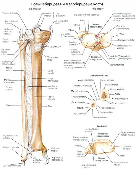
The fibula is thin and has the head of the fibula (caput fibulae) at its upper thickened (proximal) end. On the medial side of the head is the articular surface of the head of the fibula (facies articularis cdpitas fibulae) for articulation with the tibia.
The body of the fibula (corpus fibulae) is slightly curved and somewhat twisted along its longitudinal axis. The body has an anterior margin, a posterior margin, and a medial sharp interosseous margin (margo interosseus). The bone has three surfaces: lateral, posterior, and medial.

The lower and talic end of the fibula is thickened and forms the lateral malleolus (malleolus lateralis). On the medial surface of the lateral malleolus, the articular surface (facies articularis malleoli) is distinguished. For connection with the talus, behind the articular surface there is a fossa of the lateral malleolus (fossa malleoli lateralis), to which the tendons of the peroneal muscles are adjacent.
What do need to examine?
How to examine?


 [
[