Medical expert of the article

New publications
Shoulder arthroscopy
Last reviewed: 06.07.2025

All iLive content is medically reviewed or fact checked to ensure as much factual accuracy as possible.
We have strict sourcing guidelines and only link to reputable media sites, academic research institutions and, whenever possible, medically peer reviewed studies. Note that the numbers in parentheses ([1], [2], etc.) are clickable links to these studies.
If you feel that any of our content is inaccurate, out-of-date, or otherwise questionable, please select it and press Ctrl + Enter.
The shoulder complex is the most mobile of the joints in the human body. It consists of five joints: two physiological (or false) and three anatomical.
Physiological joints are the subhumeral and scapulothoracic joints, anatomical joints are the sternoclavicular, acromioclavicular and scapulohumeral joints. For the normal functioning of the shoulder complex, precise, coordinated and synchronous interaction of these joints is necessary.
What causes shoulder instability?
A large amount of information has been accumulated in the medical literature on the causes and mechanisms of occurrence of post-traumatic, recurrent shoulder dislocation; however, many authors disagree in their assessment of their role and place in the complex chain from acute traumatic shoulder dislocation to its recurrent instability. Among domestic authors, the most well-founded point of view is that of Yu.M. Sverdlov (1978), A.F. Krasnov, R.B. Akhmetzyanov (1982), D.I. Cherkes-Zade et al. (1992): they believe that the main thing in the pathogenesis of this disease is a violation of muscle balance as a result of primary traumatic dislocation, which does not respond to conservative treatment methods. Along with this, a certain significance is attached to changes in paraarticular tissues, a stretched capsule with scapulohumeral ligaments. This is the first formation on the path of the dislocating head of the humerus; the occurrence of dislocation depends on its strength and ability to withstand the pressure of the head. The cartilaginous labrum (attached to the edge of the glenoid process of the scapula) has a certain significance in the stabilization system of the shoulder joint; according to Bankart, it plays the role of a suction cup creating a "vacuum effect" between the head of the humerus and the glenoid process of the scapula (this effect significantly facilitates the rotation of the head of the humerus in the entire range of motion in the joint). Damage to the glenoid labrum leads to horizontal instability of the shoulder joint. Among domestic orthopedists, an opinion has formed about the secondary role of this damage in the pathogenesis of habitual shoulder dislocation. D.I. Cherkes-Zade et al. (1992) was the first of the domestic authors to note a very important fact: the main reason for the development of habitual shoulder dislocation and postoperative relapses is instability of the shoulder joint, caused by insufficiency of the capsular-ligamentous apparatus of the shoulder joint. Instability of the shoulder joint, as a rule, is the result of damage to several different elements of the capsular-ligamentous apparatus of the shoulder joint, each of which has a certain stabilizing function. It is obvious that in such patients it is impossible to restore the lost stability of the shoulder joint by methods that do not take into account the role of each damaged element.
Today, the theory of shoulder instability proposed by JPJon, Scott Lephart (1995) is the most modern and scientifically proven theory. Let us dwell on it in more detail.
Thus, the capsular-ligamentous structures can significantly affect stability by providing afferent feedback - reflex muscle contraction of the rotator cuff and biceps brachii in response to excessive rotation and translational movements of the humeral head. Damage to these structures leads to a significant deficit in the afferent feedback mechanism both in acute traumatic injury and in the gradual development of recurrent shoulder instability due to the combined damage to the capsular-ligamentous structures. Surgical restoration of normal anatomy of unstable joints leads to restoration of proprioceptive sensitivity.
Mechanism of injury, incidence of shoulder instability
Any healthy shoulder can dislocate if the trauma is severe enough. However, in some patients, shoulder instability can occur spontaneously, without significant trauma, due to an oversized capsule or other congenital abnormalities.
Numerous data analyzing the circumstances under which traumatic instability of the shoulder joint occurs show that displacement of the humeral head occurs in a certain position of the upper limb. Of course, the shoulder can be dislocated by direct trauma directed at the proximal part of the humerus, but an indirect, indirect force is the most common cause of anterior traumatic subluxation or dislocation. Anterior instability occurs when the shoulder is abducted above the horizontal level, at the moment of combining the forces of abduction, extension, external rotation and supination. Instability can also result from very strong muscle contraction or seizures.
The most common cause of acute traumatic instability of the shoulder is a fall with support on the hand. When the palm hits the ground, contact occurs between the upper outer part of the head of the humerus and the anteroinferior edge of the articular process of the scapula. A kind of lever is created with a fulcrum at the point of contact of the above-mentioned zones, distal to this point is the long arm of the lever, and the short arm is the most proximal part of the head of the humerus. The ratio of the lengths of these arms is 1:20, as a result of which pressure develops on the surrounding tissues at the end of the short lever, amounting to several hundred kilograms, and the bone tissue is destroyed under a force of 300 kg/cm 2. This is the most typical mechanism of shoulder dislocations, although various deviations are possible. A characteristic consequence of such an injury mechanism is significant destruction of the surrounding tissues. With such a lever mechanism, as the humeral head moves away from the center of the articular process of the scapula, the severity of the injury increases, therefore, lower dislocations are more often accompanied by bone fractures, damage to blood vessels and nerves.
The highest frequency of all shoulder joint instability is anterior instability: according to various authors, it is 75-98%.
Posterior traumatic dislocation of the shoulder is the rarest type of instability of the shoulder joint: it occurs in 2% of cases. As a rule, it is a consequence of severe direct trauma, a car accident, surgery, or electric shock treatment. With this type of instability, the head of the humerus is displaced behind the articular process of the scapula subacromially, and an impression fracture of its posterior part very often occurs. With this type of instability, diagnostic errors are most common. According to the materials of the N.N. Priorov Central Institute of Traumatology and Orthopedics, all errors were due to the fact that an X-ray examination in the axial projection was not performed.
Vertical instability of the shoulder joint was first described in 1859 by M. Meddeldorph as inferior dislocation. In its pure form, this is a very rare direction of instability. It causes severe soft tissue damage, fractures in the proximal humerus and the lower edge of the articular process of the scapula.
According to M. Wirth, superior dislocation was recorded in literature in 1834, and he also reported 12 cases. There are few mentions of this type of traumatic dislocation in modern literature: there are reports of isolated observations. The usual cause of such damage is an extreme force directed forward and upward and acting on the abducted arm. With this displacement, fractures of the acromion, acromioclavicular joint, and greater tuberosity occur. Extreme soft tissue damage occurs with the joint capsule, rotator cuff, and surrounding muscles. Neurovascular complications are usually present.
Traumatic acute and recurrent instability of the shoulder joint in patients aged 20 to 30 years occurs in 55-78% of cases during sports activities.
Traumatic instability of the shoulder joint
The earliest and most detailed description of traumatic scapulohumeral instability dates back to 460 BCE and is attributed to Hippocrates. He was the first to describe the anatomy of the shoulder joint, its types of dislocation, and the first surgical procedure he developed to reduce “that wide space into which the humeral head dislocates.” More precise descriptions of the traumatic pathology of shoulder dislocations were published in the following centuries, but the question of the “primary lesion” remains a matter of debate.
The traumatic defect that occurs in the posterolateral part of the humeral head as a result of contact with the anterior edge of the articular process of the scapula during dislocation has been identified for a long time.
In 1940, Hill and Sachs published a very clear and specific review providing information on the pathological anatomy of the humeral head in shoulder dislocations. The gist of their report is as follows.
- An impression fracture of the humeral head occurs in most shoulder dislocations.
- The longer the humeral head remains dislocated, the greater the defect.
- These impression fractures are usually larger in anteroinferior dislocations than in anterior dislocations.
- The humeral head defect is usually larger and more extensive in recurrent anterior shoulder dislocations.
Over the past decade, many authors have identified this injury arthroscopically in 82-96% of cases using large clinical data.
Moreover, the possibilities of arthroscopic surgery have allowed us to significantly deepen the morphological understanding of Bankart damage. Thanks to the work of R. Minolla, PL Gambrioli, Randelli (1995), a classification of various variants of this damage was created. Damage to the capsular-ligamentous complex of the shoulder joint with recurrent shoulder dislocation is divided into five types.
- Classic Bankart lesion - the labrum is separated from the anterior border of the glenoid process of the scapula along with the capsule and glenohumeral ligaments.
- Incomplete Bankart lesion - the labrum and capsule of the shoulder joint are not completely torn from the glenoid process of the scapula.
- The capsule is torn from the neck of the scapula, the cartilaginous labrum is torn and isolated. In this case, the capsule becomes clearly redundant, the inferior glenohumeral ligament is excessively stretched and displaced downwards. At the anterior edge of the glenoid process of the scapula, at the 2-4 o'clock position, an osteochondral lesion is determined, caused by the traumatic impact of the posterolateral part of the humeral head during the first dislocation. This is a typical, most frequent injury in recurrent anterior shoulder dislocation.
- Fracture of the anteroinferior bony rim of the articular process of the scapula, the inferior glenohumeral ligament is displaced downwards, the capsule is stretched, the cartilaginous labrum may be absent at the 2-6 o'clock position.
- Labral degeneration with anterior capsular excess. In these cases, the lesion is difficult to recognize due to cicatricial degeneration of the labrum and glenohumeral ligament complex.
Preparation
Preoperative preparation is typical for an orthopedic patient and is not specific. The operation is performed under general endotracheal anesthesia. After a comparative examination of both shoulder joints under anesthesia, the patient is placed on the operating table on the healthy side, the operated limb is fixed in a suspended state with abduction of 30° and anterior deviation of 15°, in internal rotation, with traction along the axis of the limb with a load of 5 kg on a special splint from the company "Artrex".
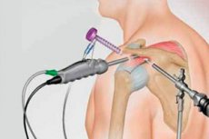
Arthroscopic stabilization of the shoulder joint
The importance of the glenohumeral ligament and labrum complex for the stable functioning of the shoulder joint is known from the works of Perthes and Bankart. In a very high percentage of cases (more than 90%) during surgical treatment of traumatic shoulder dislocation, many authors have found a rupture of these ligaments and labrum from the anteroinferior border of the glenoid process of the scapula. The inferior glenohumeral ligament functions as a primary static limiter, preventing anterior displacement of the humeral head during shoulder abduction. In addition, the labrum as an anatomical structure contributes to the formation of 25-50% of the total concavity of the relatively flat scapular socket. An intact labrum functions like the rim of a suction cup, creating a vacuum effect in the loaded shoulder, which helps the rotator cuff muscles center the humeral head in the glenoid fossa of the scapula during active range of motion. After a traumatic shoulder dislocation, the functions of the glenohumeral ligaments and labrum are lost, primarily due to the loss of their anatomical connection with the scapula.
The blood supply to the cartilaginous labrum is provided, on the one hand, by the periosteum, on the other hand, by the joint capsule. After a traumatic rupture of the cartilaginous labrum, the healing process can only begin due to the surrounding soft tissues. Fibroblastic healing is at risk in these cases. For these reasons, reconstructive measures associated with damage to these anatomical structures should primarily be aimed at their refixation to the glenoid process of the scapula as early as possible.
The surgical technique for arthroscopic treatment of shoulder instability was based on the method described by Morgan and Bodenstab for Bankart lesion repair. Arthroscopic kits from Storz and Stryker with surgical instruments from Arthrex were used for the operation.
After the surgical field has been treated and the shoulder joint landmarks have been marked on the skin, the shoulder joint is punctured with a syringe and a puncture needle from the posterior approach in the direction of the medial part of the apex of the coracoid process of the scapula. The needle entering the shoulder joint is felt as a slight "gap", after which synovial fluid begins to flow from the needle. Next, 50-60 ml of physiological solution for its joint cavity is injected into the joint cavity. After this, a 0.5 cm long skin incision is made in the projection of the posterior approach. Through it, using a blunt trocar, repeating the direction of the puncture needle, an arthroscope case is inserted into the joint, the trocar is changed to an optical arthroscope with a video camera. Through the anterior approach, located between the apex of the coracoid process and the head of the humerus, a plastic cannula is inserted into the joint along the guide wire to drain fluid from the joint. The necessary arthroscopic instruments are inserted into the joint through this cannula, after which diagnostic arthroscopy of the shoulder joint is performed using a standard 30-degree arthroscope with a diameter of 4 mm.
The fluid is injected into the joint through the arthroscope casing using a mechanical pump (to maintain constant pressure of the saline solution in the joint). Experience shows that the use of a mechanical pump is safe and helps the surgeon to constantly monitor possible tissue bleeding. After a Bankart lesion has been visually diagnosed (tear of the anteroinferior part of the cartilaginous labrum with the middle and lower glenohumeral ligaments and the capsule of the shoulder joint from the articular process of the scapula, sometimes with a bone fragment), the degree of mobility and depth of separation of the soft tissues from the scapular edge and neck are determined using a search hook.
When the detachment of the cartilaginous labrum is small, it must be increased using a special manual raspatory.
Next, an electric rotary burr is inserted into the joint through a plastic cannula to treat the bone surface (arthroshaver), with its help the entire anterior edge of the articular process of the scapula is treated up to the bleeding bone wound.
This stage is very important, as it creates conditions for fibroblastic healing between the Bankart lesion and the glenoid process of the scapula. Particular attention should be paid to the careful, uniform treatment of the bone surface, so as not to damage the articular cartilage and not to disrupt the spherical surface of the glenoid process of the scapula. When pinpoint bleeding from the bone is obtained, the depth of treatment is considered sufficient.
The detached scapulohumeral complex (inferior glenohumeral ligament + labrum) is captured with a special clamp-guide, moved to the anatomical attachment site on the articular process of the scapula and held in this position.
The next very important stage is the application of transglenoid sutures. A pin with an eye (30 cm long, 2 mm in diameter) is inserted through the guide clamp, the cartilaginous labrum is pierced, and the entire complex is shifted as upward as possible (cranially) by 5-10 mm. This is a very important moment of physiological tension of the inferior glenohumeral ligament and its fixation in the anatomical attachment site on the anterior edge of the glenoid process of the scapula. In this case, the pin should pass 2-3 mm below the edge of the glenoid process, through the neck of the scapula at an angle of 30° and 10-15° medially to the glenoid plane. The pin is inserted using a drill, the sharp end of the pin comes out through the posterior surface of the neck of the scapula and the infraspinatus muscle under the skin. A 1 cm long incision is made with a scalpel, and the sharp end of the pin is inserted into it. The exit point of the needle on the scapular surface is preliminarily determined using a stereoscopic arc, which is fixed on the base of the guide clamp, which helps to avoid accidental damage to the suprascapular nerve (n. suprascapularis). A monofilament suture thread "polydioxanone" No. 1 is inserted into the eye of the needle. Removing the needle by the sharp end, the suture thread is passed through the soft tissue complex and the neck of the scapula. The second needle is passed in a similar manner 1 cm higher (cranial) than the first, the free end of the first thread is tied in its eye, and the second thread is tied to it. When passing through the scapula, the threads are brought out into a skin incision 1 cm higher than the first. The ends of the first thread are tied together under the fascia of the subscapularis muscle when traction is removed from the limb and the arm is given a position of adduction and internal rotation.
A total of 3-4 similar sutures are applied, placed sequentially from bottom to top. The sutures securely fix the cartilaginous labrum on the glenoid process of the scapula in an anatomical position. In this case, the restored complex of the scapulohumeral ligaments and cartilaginous labrum should look like a stretched structure, and the labrum should be located above the anterior edge of the glenoid process of the scapula, evenly along the entire perimeter.
The skin wounds are sutured and an aseptic bandage is applied. The limb is fixed in internal rotation in an immobilization splint.
Thus, the basic operating principle of the arthroscopic Bankart suture for primary or recurrent posttraumatic instability of the shoulder joint is anatomically sound refixation of the glenoid labrum with the lig. glenohumerale complex to the anterior edge of the glenoid process of the scapula. After arthroscopic refixation, the labrum can again function as an attachment site for these ligaments and as a sealing ring between the glenoid process of the scapula and the head of the humerus, providing a suction effect due to negative pressure in this space throughout the entire range of motion in the shoulder joint.
 [
[