Medical expert of the article

New publications
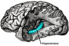
Hippocampus
Last reviewed: 04.07.2025

All iLive content is medically reviewed or fact checked to ensure as much factual accuracy as possible.
We have strict sourcing guidelines and only link to reputable media sites, academic research institutions and, whenever possible, medically peer reviewed studies. Note that the numbers in parentheses ([1], [2], etc.) are clickable links to these studies.
If you feel that any of our content is inaccurate, out-of-date, or otherwise questionable, please select it and press Ctrl + Enter.
If ancient Greek mythology called the Hippocampus the lord of fish, representing it in the form of a sea monster - a horse with a fish tail, then the hippocampus of the brain, which is its important structure, received this name due to the similarity of its shape in the axial plane with an unusual needle-shaped fish of the genus Hippocampus - a seahorse.
By the way, the second name of the curved internal structure of the temporal lobe of the brain, given to it by anatomists in the middle of the 18th century – Ammon’s horn (Cornu Ammonis), is associated with the Egyptian god Amun (in the Greek form – Ammon), who was depicted with ram’s horns.
The structure of the hippocampus and its structures
The hippocampus is a complex structure deep within the temporal lobe of the brain, between its medial side and the inferior horn of the lateral ventricle, forming one of its walls.
The elongated interconnected structures of the hippocampus (folds of gray matter of the archicortex that are folded into each other) are located along the longitudinal axis of the brain, one in each of the temporal lobes: the right hippocampus and the contralateral left hippocampus. [ 1 ]
In adults, the size of the hippocampus—the length from the front to the back—varies between 40 and 52 mm.
The main structures are the hippocampus itself (Cornu Ammonis) and the dentate gyrus (Gyrus dentatus); specialists also distinguish the subicular cortex, which is an area of gray matter of the cerebral cortex surrounding the hippocampus. [ 2 ]
The horn of Ammon forms an arch, the rostral (anterior) part of which is enlarged and is defined as the head of the hippocampus, which curves backward and downward, forming on the medial side of the temporal lobe the hook of the hippocampus or uncus (from the Latin uncus - hook) - (Uncus hippocampi). Anatomically, it is the anterior end of the parahippocampal gyrus (Gyrus parahippocampi), which is curved around the hippocampus itself and protrudes into the floor of the temporal (lower) horn of the lateral ventricle.
Also in the rostral part there are thickenings in the form of three or four separate protrusions of the cortical convolutions, which are called the hippocampal fingers (Digitationes hippocampi).
The middle part of the structure is defined as the body, and the part of it called the alveus is the floor of the lateral ventricle (temporal horn) of the brain and is almost entirely covered by the choroid plexus, which is a combination of the pia mater and ependyma (tissue lining the cavity of the ventricles). The fibers of the white matter of the alveus are collected in thickened bundles in the form of a fringe or fimbria (Fimbria hippocampi), then these fibers pass into the fornix of the brain.
Below the hippocampus is its main outlet, the superior flat part of the parahippocampal gyrus, called the subiculum. This structure is separated by a shallow rudimentary fissure or groove of the hippocampus (Sulcus hippocampalis), which is a continuation of the groove of the corpus callosum (Sulcus corporis callosi) and runs between the parahippocampal and dentate gyri. [ 3 ]
The dentate gyrus of the hippocampus, also called the parahippocampus, is a three-layered concave groove separated from the fibril and subiculum by other grooves.
It should also be borne in mind that the hippocampus and the adjacent dentate and parahippocampal gyri, subiculum and entorhinal cortex (part of the cortex of the temporal lobe) form the hippocampal formation - in the form of a bulge at the bottom of the temporal horn of the lateral ventricle.
In this zone – in the medial surfaces of both hemispheres of the brain (Hemispherium cerebralis) – a set of brain structures that are part of the limbic system of the brain are localized. The limbic system and the hippocampus, as one of its structures (along with the amygdala, hypothalamus, basal ganglia, cingulate gyrus, etc.), are connected not only anatomically, but also functionally. [ 4 ]
The hippocampus is supplied with blood by the vessels that supply the temporal lobes of the brain, that is, by the branches of the middle cerebral artery. In addition, blood enters the hippocampus through the branches of the posterior cerebral artery and the anterior choroidal artery. And the outflow of blood goes through the temporal veins - the anterior and posterior.
Neurons and neurotransmitters of the hippocampus
The heterogeneous cortex of the hippocampus - allocortex - is thinner than the cerebral cortex and consists of a superficial molecular layer (Stratum molecular), a middle layer Stratum pyralidae (consisting of pyramidal cells) and a deep layer of polymorphic cells.
Depending on the features of the cellular structure, Ammon's horn is divided into four different areas or fields (the so-called Sommer sectors): CA1, CA2, CA3 (the area of the hippocampus itself, covered by the dentate gyrus) and CA4 (in the dentate gyrus itself).
Together, they form a neural trisynaptic circuit (or circuit), in which the functions of transmitting nerve impulses are performed by hippocampal neurons, in particular: excitatory pyramidal neurons of the CA1, CA3 and subiculum fields, characteristic of the structures of the anterior parts of the brain. Glutamatergic pyramidal neurons, which have dendrites (afferent processes) and axons (efferent processes), are the main type of cells in the nervous tissue of the hippocampus.
In addition, there are stellate neurons and granule cells concentrated in the granule cell layer of the dentate gyrus; GABAergic interneurons - multipolar intercalary (association) neurons of the CA2 field and parahippocampus; basket (inhibitory) neurons of the CA3 field, as well as the recently identified intermediate OLM interneurons in the CA1 region. [ 5 ]
Chemical messengers that are released from the secretory vesicles of the principal cells of the hippocampus into the synaptic cleft to transmit nerve impulses to target cells – neurotransmitters or neuromediators of the hippocampus (and the entire limbic system) – are divided into excitatory and inhibitory. The former include glutamate (glutamic acid), norepinephrine (norepinephrine), acetylcholine and dopamine, the latter – GABA (gamma-aminobutyric acid) and serotonin. Depending on which neurotransmitters act on the transmembrane nicotinic (ionotropic) and muscarinic (metabotropic) receptors of the neural circuits of the hippocampus, the activity of its neurons is excited or suppressed. [ 6 ]
Functions
What is the hippocampus of the brain responsible for, what functions does it perform in the central nervous system? This structure is connected to the entire cerebral cortex by indirect afferent pathways passing through the entorhinal cortex and subiculum and is involved in the processing of cognitive and emotional information. To date, the best known is how the hippocampus and memory are connected, and researchers are also finding out how the hippocampus and emotions are connected.
Neuroscientists studying the functions of the hippocampus have divided it topographically into the posterior or dorsal part and the anterior or ventral part. The posterior part of the hippocampus is responsible for memory and cognitive functions, and the anterior part is responsible for the manifestation of emotions. [ 7 ]
It is believed that information is sent from multiple sources via commissural nerve fibers (commissures) of the temporal lobe cortex to the hippocampus, which it encodes and integrates. From short-term memory [ 8 ] it forms long-term declarative memory (about events and facts) due to long-term potentiation, that is, a special form of neural plasticity - an increase in neuronal activity and synaptic strength. The retrieval of information about the past (memories) is also regulated by the hippocampus. [ 9 ]
In addition, the hippocampal structures participate in the consolidation of spatial memory and mediate spatial orientation. This process consists of cognitive mapping of spatial information, and as a result of its integration in the hippocampus, mental representations of the location of objects are formed. And for this, there is even a special type of pyramidal neurons - place cells. Presumably, they also play an important role in episodic memory - recording information about the environment in which certain events occurred. [ 10 ]
As for emotions, the most important of the cerebral structures that are directly related to them is the limbic system and its integral part – the hippocampal formation. [ 11 ]
And in this regard, it is necessary to explain what the hippocampal circle is. This is not an anatomical structure of the brain, but the so-called medial limbic chain or emotional circle of Papez. Considering the hypothalamus to be the source of human emotional expression, the American neuroanatomist James Wenceslas Papez in the 1930s put forward his concept of the path of formation and cortical control of emotions and memory. In addition to the hippocampus, this circle included the mammillary bodies of the base of the hypothalamus, the anterior nucleus of the thalamus, the cingulate gyrus, the cortex of the temporal lobe surrounding the hippocampus, and some other structures. [ 12 ]
Further studies have clarified the functional connections of the hippocampus. In particular, the amygdala (Corpus amygdaloideum), which is located in the temporal lobe (in front of the hippocampus), was recognized as the emotional center of the brain responsible for the emotional evaluation of events, the formation of emotions, and making emotional decisions. As part of the limbic system, the hippocampus and amygdala/amygdala act together in stressful situations and when a feeling of fear arises. The parahippocampal gyrus is also involved in negative emotional reactions, and the consolidation of emotionally expressed (scary) memories occurs in the lateral nuclei of the amygdala. [ 13 ]
The hypothalamus and hippocampus, located in the midbrain, have numerous synaptic connections, which determines their participation in the response to stress. Thus, the anterior part of the hippocampus, providing negative feedback, controls stress reactions of the functional neuroendocrine axis hypothalamus-pituitary-adrenal cortex. [ 14 ]
In search of an answer to the question of how the hippocampus and vision are connected, neuropsychological studies have established the involvement of the parahippocampal gyrus and perirhinal cortex (part of the cortex of the medial temporal lobe) in visual recognition of complex objects and memorization of objects.
And what connections the hippocampus and the olfactory brain (Rhinencephalon) have is known exactly. Firstly, the hippocampus receives information from the olfactory bulb (Bulbus olfactorius) – through the amygdala. Secondly, the hook of the hippocampus (uncus) is the olfactory center of the cerebral cortex and can be attributed to the rhinencephalon. Thirdly, the cortical area responsible for olfaction also includes the parahippocampal gyrus, which stores information about odors. [ 15 ] Read more – Olfaction
Hippocampal diseases and their symptoms
Experts consider the hippocampus to be a fairly vulnerable brain structure; damage to it (including traumatic brain injuries) and associated diseases can cause various symptoms – neurological and mental.
Modern neuroimaging methods help to identify morphometric changes in the hippocampus (its volume), which occur with hypoxic damage and certain diseases of the brain, as well as with its reduction deformations.
An important clinical sign is considered to be hippocampal asymmetry, since, presumably, the left and right hippocampus are affected differently with aging. According to some studies, the left hippocampus plays a major role in episodic verbal memory (speech reproduction of memories), and the right hippocampus plays a major role in the consolidation of spatial memory. According to measurements, in people over 60 years of age, the difference in their volumes is 16-18%; with age, it increases, and in men, compared to women, the asymmetry is more pronounced. [ 16 ]
A slight shrinkage of the hippocampus that occurs with age is considered normal: atrophic processes in the medial temporal lobe and entorhinal cortex begin to occur closer to the age of seventy. But a significant reduction in the size of the "seahorse" of the brain increases the risk of developing dementia, the early symptoms of which are manifested by short episodes of memory loss and disorientation. Read more in the article - Symptoms of dementia
The reduction of the hippocampus is much more pronounced in Alzheimer's disease. However, it is still unclear whether this is a result of this neurodegenerative disease or serves as a prerequisite for its development. [ 17 ]
According to research, patients with generalized depressive disorder and post-traumatic stress disorders have bilateral and unilateral reduction in hippocampal volume by 10-20%. Long-term depression is also accompanied by a decrease or disruption of neurogenesis in the hippocampus. [ 18 ] According to neurophysiologists, this occurs due to increased levels of cortisol. This hormone is intensively produced and released by the adrenal cortex in response to physical or emotional stress, and its excess negatively affects the pyramidal neurons of the hippocampus, impairing long-term memory. It is because of high levels of cortisol that the hippocampus decreases in patients with Itsenko-Cushing's disease. [ 19 ], [ 20 ]
- Read also – Symptoms of stress
A reduction in the number or alteration of hippocampal nerve cells may also be associated with inflammatory processes (neuroinflammation) in the temporal lobe of the brain (for example, in bacterial meningitis, in encephalitis caused by the herpes simplex virus type I or II) and long-term activation of microglia, whose immune cells (macrophages) release proinflammatory cytokines, proteinases and other potentially cytotoxic molecules.
The volume of this cerebral structure may decrease in patients with brain gliomas, since tumor cells produce the neurotransmitter glutamate into the extracellular space, the excess of which leads to the death of hippocampal neurons.
In addition, a number of studies with MRI volumetry of the hippocampus have recorded its reduction in traumatic brain injury, epilepsy, mild cognitive impairment, Parkinson's and Huntington's diseases, schizophrenia, Down and Turner syndromes. [ 21 ]
Insufficient nutrition of the nervous tissue – hippocampal hypotrophy – may have an ischemic etiology after strokes; in drug addiction, in particular opioid addiction, hypotrophy is observed due to disturbances in the metabolism of dopamine by psychoactive substances.
Disorders caused by a deficiency of certain elements affect the trophism of the nervous tissue of the entire hippocampal formation, negatively affecting the functioning of the central nervous system. Thus, vitamin B1 or thiamine and the hippocampus are linked by the fact that in cases of chronic deficiency of this vitamin, the processes of formation of short-term memory are disrupted. It turned out that with a deficiency of thiamine (the risk of which is increased in alcoholics) in the dentate gyrus and hippocampal fields CA1 and CA3, the number of pyramidal neurons and the density of their afferent processes can decrease, which is why there are failures in the transmission of nerve impulses. [ 22 ], [ 23 ] Long-term thiamine deficiency can cause Korsakov's syndrome.
Progressive decrease in the volume of nervous tissue with loss of neurons – hippocampal atrophy – occurs in almost the same diseases, including Alzheimer's and Itsenko-Cushing's diseases. Risk factors for its development are considered to be cardiovascular diseases, depression and stress conditions, epileptic status, diabetes mellitus, arterial hypertension, [ 24 ] obesity. And symptoms include memory loss (in Alzheimer's disease – up to anterograde amnesia ), [ 25 ], [ 26 ] difficulties in performing familiar processes, spatial definition and verbal expression. [ 27 ]
In case of disruption of the structural organization of cells of the fields of Ammon's horn and the subiculum area and loss of some pyramidal neurons (atrophy) - with expansion of the interstitium and proliferation of glial cells (gliosis) - sclerosis of the hippocampus is determined - mesial sclerosis of the hippocampus, mesial temporal or mesial temporal sclerosis. Sclerosis is observed in patients with dementia (causing loss of episodic and long-term memory), and also leads to temporal epilepsy. [ 28 ] Sometimes it is defined as limbic temporal or hippocampal, that is, epilepsy of the hippocampus. Its development is associated with the loss of inhibitory (GABAergic) interneurons (which reduces the ability to filter afferent signals of the entorhinal cortex and leads to hyperexcitability), disruption of neurogenesis and proliferation of axons of the granular cells of the dentate villin. Additional information in the article - Epilepsy and epileptic seizures - Symptoms
As clinical practice shows, hippocampal tumors are rarely found in this cerebral structure, and in most cases it is a ganglioglioma or dysembryoplastic neuroepithelial tumor - a slowly growing benign glioneuronal neoplasm consisting mainly of glial cells. Most often it occurs in childhood and young age; the main symptoms are headache and difficult to treat chronic seizures.
Congenital anomalies of the hippocampus
In such malformations of the cerebral cortex as focal cortical dysplasia, hemimegalencephaly (unilateral enlargement of the cerebral cortex), schizencephaly (the presence of abnormal cortical clefts), polymicrogyria (reduction of convolutions), as well as periventricular nodular heterotopia accompanied by seizures and visual-spatial disorders, a decrease in the hippocampus is noted.
Abnormal enlargement of the amygdala and hippocampus has been found by researchers in the presence of early infantile autism syndrome. Bilateral enlargement of the hippocampus is observed in children with lissencephaly of the brain, abnormal thickening of the convolutions (pachygyria) or with subcortical laminar heterotopia - doubling of the cerebral cortex, the manifestation of which are epileptic seizures. More information in the materials:
Associated with underdevelopment of the brain, hypoplasia of the hippocampus and often of the corpus callosum is found in newborns with severe encephalopathy with a mutation in the WWOX gene, which codes for the enzyme oxidoreductase. This congenital anomaly, leading to early death, is characterized by the absence of spontaneous movements in the infant and the absence of a response to visual stimuli, as well as seizures (which appear several weeks after birth).
Hippocampal inversion – a change in its anatomical position and shape – also represents a defect in the intrauterine development of the hippocampus itself (Cornu Ammonis), the formation of which from the folds of the gray matter of the archicortex is completed by the 25th week of pregnancy.
Incomplete hippocampal inversion, also known as hippocampal malrotation or hippocampal inversion with malrotation, is the formation of a spherical or pyramidal hippocampus, which is most often observed in the left temporal lobe – with a decrease in size. Morphological changes in the adjacent sulci may be observed. The anomaly is detected in patients with and without seizures, with and without other intracranial defects.
A hippocampal cyst is also a congenital anomaly – a small cavity filled with cerebrospinal fluid (a dilated perivascular space limited by a thin wall) of a round shape. Residual hippocampal cysts, synonym – remnant sulcus cysts (Sulcus hippocampalis), are formed during incomplete involution of the embryonic cleft of the hippocampus during intrauterine development. The characteristic localization of cysts is on the side at the top of the hippocampal sulcus, between the Cornu Ammonis and Gyrus dentatus. They do not manifest themselves in any way and are most often discovered accidentally during routine MRI examinations of the brain. According to some data, they are detected in almost 25% of adults.
The Hippocampus and Coronavirus
Since the onset of the spread of covid-19, doctors have noted forgetfulness, anxiety, and depression in many recovered patients, and often hear complaints of “brain fog” and increased irritability.
The coronavirus that causes covid-19 is known to enter cells through receptors in the olfactory bulb (Bulbus olfactorius), which manifests itself as a symptom known as anosmia or loss of smell.
The olfactory bulb is connected to the hippocampus, and according to neurodegenerative disease researchers at the Alzheimer's Association, damage to it is responsible for the cognitive impairment seen in covid-19 patients, particularly problems with short-term memory.
There was a recent announcement that a large-scale study of the effects of coronavirus on the brain and the causes of cognitive decline will soon begin, involving scientists from nearly four dozen countries – under the technical guidance and coordination of the WHO.
Read also: Coronavirus lingers in the brain even after recovery
Diagnosis of hippocampal diseases
The main methods for diagnosing diseases associated with certain damage to the structures of the hippocampus include examination of the neuropsychic sphere, magnetic resonance imaging and computed tomography of the brain.
Physicians prefer to visualize the hippocampus with MRI: with standard T1-weighted sagittal, coronal, diffusion-weighted axial images, T2-weighted axial images of the whole brain, and T2-weighted coronal images of the temporal lobes. To detect pathological changes in the fields of the hippocampus itself, the dentate or parahippocampal gyrus, MRI at 3T is used; MRI with a higher field may also be required. [ 29 ]
Also performed: Doppler ultrasonography of the cerebral vessels, EEG – encephalography of the brain.
Details in the publications:
Treatment of hippocampal diseases
Congenital anomalies of the hippocampus associated with underdevelopment and reduction deformations of the brain cannot be cured: children are doomed to disability due to cognitive impairment of varying degrees of severity and associated behavioral disorders.
How to treat some of the diseases listed above, read in the publications:
- Epilepsy - Treatment
- Alzheimer's Dementia - Treatment
- New Treatments for Alzheimer's Disease
- Treatment of depression
- Vitamins for the brain
In cases where anticonvulsants, that is, antiepileptic drugs, do not cope with attacks in mesial temporal lobe epilepsy, [ 30 ] they resort to the last resort - surgical treatment.
The surgeries include: hippocampectomy – removal of the hippocampus; limited or extended epileptogenic zone ectomy (resection or excision of affected structures); temporal lobectomy with preservation of the hippocampus; selective resection of the hippocampus and amygdala (amygdalohippocampectomy). [ 31 ]
According to foreign clinical statistics, in 50-53% of cases after surgery, epileptic seizures in patients stop; 25-30% of those operated on have seizures 3-4 times a year.
How to train the hippocampus?
Since the hippocampus (its dentate gyrus) is one of the few cerebral structures where neurogenesis or neural regeneration (the formation of new neurons) occurs, the process of memory deterioration (provided that the underlying disease is treated) can be positively influenced by exercise.
Aerobic exercise and any moderate physical activity (especially in old age) have been shown to promote neuronal survival and stimulate the formation of new hippocampal nerve cells. Incidentally, exercise reduces stress and improves depression. [ 32 ], [ 33 ]
In addition, cognitive stimulation, that is, mental exercises, helps train the hippocampus: memorizing poems, reading, solving crosswords, playing chess, etc.
How to increase the hippocampus, because in old age it becomes smaller? A method proven by researchers is physical exercises, thanks to which the perfusion of the hippocampus increases, and the formation of new cells of the nervous tissue is more active.
How to restore the hippocampus after stress? Do mindfulness meditation, which is a mind training practice aimed at slowing down racing thoughts, releasing negativity, and achieving calmness for the mind and body. As the results of a study by one of the East Asian universities have shown, meditation helps to reduce the level of cortisol in the blood.