Medical expert of the article

New publications
Dermatophytosis
Last reviewed: 29.06.2025

All iLive content is medically reviewed or fact checked to ensure as much factual accuracy as possible.
We have strict sourcing guidelines and only link to reputable media sites, academic research institutions and, whenever possible, medically peer reviewed studies. Note that the numbers in parentheses ([1], [2], etc.) are clickable links to these studies.
If you feel that any of our content is inaccurate, out-of-date, or otherwise questionable, please select it and press Ctrl + Enter.
A widespread superficial fungal lesion of keratinized tissues - the stratum corneum of the epidermis, hair and nails - caused by specific filamentous dermatophyte fungi and defined as dermatophytosis (from Greek dermatos - skin and phyton - plant), as well as epidermophytosis, dermatophytosis or dermatomycosis (from Greek dermatos - skin and mykes - fungus). [1]
Epidemiology
The World Health Organization estimates the worldwide incidence of dermatophytosis at 10,000-15,000 for every 100,000 people.
Dermatophytoses, as common superficial fungal infections worldwide, are more common in tropical and subtropical countries like India due to high humidity and ambient temperature. Increased urbanization, closed shoes and tight clothing also predispose to higher prevalence. [2]
It is estimated that superficial fungal infections affect approximately 20-25% of the world's population. In Brazil, studies by Siqueira et al (2006) and Brilhante et al (2000) [4] showed that the prevalence of dermatophytosis among cutaneous lesions ranged from 18.2% to 23.2%. [5]
In Europe, the zoophilic dermatomycete Microsporum canis is the most frequent cause of scalp dermatophytosis in the Mediterranean, Hungary, Poland, Austria and Germany. More than 85% of dermatologists' patients are children and adolescents.
And nearly 14% of U.S. Adults, more than 16% of French adults, about 8% of Canadians and 3% of Britons have onychomycosis.
Causes of the dermatophytoses
Among dermatophytes (i.e. Anthropophytes) parasitizing human skin, the main causative agents of epidermophytosis or dermatophytosis are recognized as microscopic fungi of the genus Trichophyton (trichophyton) of the family Arthrodermataceae and representatives of the same family: Microsporum (Microsporum) and Epidermophyton (Epidermophyton). [7]
Red Trichophyton trichophyton rubrum, the most common human dermatophyte (dermatomycete), is the cause of dermatophytosis, which is called trichophytosis, trichomycosis, rubrophytosis, or rubromycosis.
If the cause of the skin lesion microsporum, the fungal disease, also by a specific pathogen, is most often called microsporia. So in terms of the etiology of the skin lesion, microsporia and dermatophytosis are synonymous.
And by localization of the lesion are synonymous caused by tricho- and epidermophyton dermatophytosis of the nails and onychomycosis (from Greek onychos - nail and mykes - fungus).
Thus, depending on the causative agent, such types of dermatophytosis are distinguished as:
- Trichophytosis (fungal diseases of the skin, hair, and nails);
- Microsporia (dermatomycoses of the skin and hair);
- Epidermophytosis (affects the skin of the feet, skin folds, and nails).
Separately distinguish favus (parsha) - a chronic scarring form of dermatomycosis of the head, caused by the anthropophilic fungus Trichophyton schoenleinii, discovered by the German physician Johann Schoenlein (1793-1864).
Risk factors
Risk factors for the development of dermatophytosis include xerosis (dry skin), immunosuppression, obesity, diabetes mellitus, skin trauma, high ambient temperature and humidity levels, excessive sweating, and lack of proper hygiene.
Is dermatophytosis contagious? Yes, dermatophyte fungi can be transmitted through direct contact with an infected person or animal, as well as indirect contact through towels, clothing, hats, shoes and other household items. [8] Other epidemiological studies confirm the high frequency of onychomycosis in relation to other forms of ringworm. [9], [10] This may be attributed to increased use of swimming pools, increased participation in sports, wearing closed toed shoes in both professional and leisure settings, and increased incidence of diabetes and vascular disease. [11], [12]
Dermatophytosis can easily be contracted through contact with viable fungal spores in places such as swimming pools, saunas, public showers, nail salons, gyms, etc.
Pathogenesis
Dermatophytes are hyaline filamentous molds consisting of mycelium (absorbing nutrients) and capable of forming spores (conidia). They are keratinophilic fungi, and the pathogenesis of dermatophytosis is due to their keratinolytic properties. These fungi do not attack mucosal surfaces, but target the keratin of the skin and its appendages, as this structural fibrillar protein is essential for their nutrition and growth.
With their special spores (arthroconidia), dermatophytes attach to the epidermis and begin to germinate in the stratum corneum. And fungi that "specialize" in hair penetrate the ectotrix (outer hair shaft) and the core of the hair shaft (endotrix).
In doing so, they hide components of their cell wall from the human immune system, inhibiting T lymphocytes and suppressing the immune response.
When arthroconidia begin to sprout into the stratum corneum of the epidermis, germ tubes are formed that promote the spread of infection. And the proteolytic enzymes produced by the fungi break down keratinized tissue into oligopeptides and free amino acids, which are used as nutrients.
In addition, as a result of metabolism of released amino acids there is a release of ammonia, changing the pH of the skin from acidic to alkaline, which creates conditions for increased activity of enzymes of dermatophytes and increased proteolytic degradation of keratin of the stratum corneum of the skin, hair and nail plates.
Symptoms of the dermatophytoses
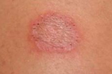
The main symptoms of dermatophytosis include rash, scaling, and itching, and the first signs appear as erythematous scaly nodules that gradually transform into annular or round red patches or plaques with lucency in the center and scaling at the edges. [13] Rashes may be located on the scalp, neck, trunk, extremities, and groin. Clinical types of dermatophyte infection are usually defined by the localization of the lesions.
Inguinal dermatophytosis
Dermatophytosis inguinalis or inguinal epidermophytosis - with blistering red flaky patches with raised borders - affects the skin of the inner upper thighs and can spread to the buttocks and abdomen.
Dermatophytosis inguinale is more common in men than in women. Also see - the pathogen of inguinal epidermophytosis (Epidermophyton floccosum)
Dermatophytosis under the breasts can occur in women, for more information see. - mycosis of large folds
Dermatophytosis of the scalp
This fungal disease develops when infected with the dermatophytes Microsporum canis (transmitted from pets - dogs and cats), Microsporum ferrugineum and Trichophyton tonsurans (transmitted from humans). Most often, dermatophytosis in children occurs on the scalp (and is traditionally called ringworm). When the cause is associated with Tr. Tonsurans (in Latin tonsurans - shaving) on the scalp appear multiple spots covered with scales and devoid of hair, and when affected by microsporums, a large single alopetic (hairless) spot with more pronounced inflammation of the skin in the form of redness and itching is formed. Scattered broken hairs of varying lengths or areas of discrete hair loss in the form of dark dots may be observed in the affected areas.
Other dermatophytes also affect the scalp: Microsporum audouinii, Microsporum gypseum, Trichophyton mentagrophytes, Trichophyton violaceum, Trichophyton schoenleinii, Trichophyton verrucosum, Trichophyton soudanense and Trichophyton rubrum.
In the case of parsha (phavus), there are many yellowish, round crusted, grouped spots with tufts of hair protruding in their center. A "mouse odor" is often present. Growth continues for several months, after which the crusts fall off, leaving a shiny bare area without hair. It is chronic and can last for years.
Dermatophytosis of the feet
This localized dermatomycosis, often referred to as athlete's foot, can be caused by the fungi Epidermophyton floccosum, Trichophyton rubrum and the causative agent of epidermophytosis of the feet (Trichophyton interdigitale); in the latter case, the most common form, interdigitale dermatophytosis, develops, with symptoms such as cracking, maceration, peeling and itching of the skin between the toes.
In plantar dermatomycosis, the soles, heels and sides of the foot are affected - with redness, flaking and gradual thickening of the stratum corneum of the epidermis. Dyshidrotic dermatophytosis - the dyshidrotic form of foot dermatomycosis is manifested by marked inflammation with the formation of blisters and vesicles. Read more - epidermophytosis of the feet
Dermatophytosis of the hands
All the details are in the publications:
Dermatophytosis of smooth skin
The dermatophytes Trichophyton rubrum, Trichophyton mentagrophytes and Epidermophyton floccosum can affect the smooth skin of the trunk, extremities or face. Thus, trunk dermatophytosis presents as one or many flaky, ring-shaped lesions with unaffected skin in the middle, slightly raised red borders and a distinct border between the affected and unaffected skin. Blisters or follicular nodules may form on the border of the affected areas. Itching may be absent.
Complications and consequences
In the acute form of fungal invasion, some patients may experience a delayed-type hypersensitivity reaction, also known as a trichophytosis reaction, which is manifested by general malaise, fever, headache and muscle pain.
And the extreme local immunologic response to fungal infection of the scalp hair follicles is kerion, a large soft mass on the scalp with blisters and crusts and can lead to destruction of hair follicles and scarring alopecia.
Also a complication of advanced dermatophytosis is cracking of the skin, which can lead to the attachment of bacterial infection and inflammation of deeper layers of the skin.
Severe itching in foot dermatophytosis is complicated by deep scratching, causing excoriations (open wounds) that are also susceptible to bacterial infection. In addition, scratching infected areas can result in the spread of the fungus to other parts of the body.
Although these dermatoses are not serious in terms of mortality or psychological morbidity, they have serious clinical consequences, causing chronic, difficult-to-treat skin lesions. Moreover, they lead to a reduced quality of life for patients and cause disfigurement, which affects self-esteem and vanity and may even lead to social discrimination. [14]
Diagnostics of the dermatophytoses
Even to an experienced dermatologist, a visual examination of the patient and patient history are not enough to detect trichophytosis, microsporia, or epidermophytosis.
Instrumental diagnostics including dermatoscopy, Wood's lamp detection of fungal infection, and reflectance confocal microscopy are performed.
To identify dermatophyte infections, the following tests are required: scraping of the affected area for microscopic examination for fungi; treatment of samples of affected hair or skin scales with potassium hydroxide (KOH) [15], [16], [17] and seeding - culture of fungi.
PCR (polymerase chain reaction) is increasingly being used as a diagnostic assay for the detection of dermatophytes, which is a more sensitive method because it can detect dermatophyte DNA. [18] Uniplex PCR is useful for direct detection of fungi in clinical specimens with sensitivity and specificity of 80.1% and 80.6%, respectively, compared to culture. [19] Multiplex PCR for fungal detection of dermatophytes detects 21 dermatomycosis pathogens with DNA detection by agarose gel electrophoresis.
Differential diagnosis
Differential diagnosis is designed to distinguish dermatophytosis from other causative agents of superficial mycoses (in particular, keratomycosis), as well as from flat fever, allergic contact dermatitis, seborrheic dermatitis, dyshidrotic and coin-like eczema, psoriasis, discoid cutaneous lupus erythematosus, sycosiform atrophic folliculitis.
Who to contact?
Treatment of the dermatophytoses
The treatment of dermatophytosis is usually a long and laborious process. The choice of adequate treatment is determined by the site and extent of the lesion, the type of fungus affected, and the efficacy, safety profile and pharmacokinetics of available antifungal agents. [20]
First-line therapy is based on the use of topical agents, usually the antifungal imidazoles. 15 If this therapy is ineffective, oral therapy with antifungal agents such as terbinafine, itraconazole, ketoconazole, and fluconazole usually follows. [21] Combination therapy with topical and oral antifungal and anti-inflammatory agents has been used in an attempt to increase cure rates.
Systemic therapy is indicated when lesions are generalized, recurrent, chronic, or unresponsive to topical therapy. Conventional oral regimens are associated with long treatment duration and poor adherence. [23]
Topical antifungal drugs (antimycotics) are prescribed to treat dermatophytosis. The main medicines are antifungal agents:
Ointment terbinafine (Terbizil, Terbized, Lamisil, Lamifen), Sertaconazole (Zalain ), Miconazole, Econazole, etc.; nail cream and varnish batrafen (with cyclopyroxa olamine). For more information see:
Griseofulvin, Ketoconazole, Fluconazole and other pills for skin fungus are always used in the systemic therapy of scalp dermatophytosis.
Several comparative studies have shown that fluconazole was the least active of the antifungal agents evaluated, with its effect varying according to the species of pathogen. [24], [25]
In superficial fungal lesions of keratinized tissues, herbal treatment with such medicinal plants as peppermint, St. John's wort, garlic, medicinal ginger, common oregano, soursop triangular, mirena dye, centella asiatica can be auxiliary. In addition, some folk remedies such as apple cider vinegar, baking soda, and lemon juice can also help in the treatment.
More useful information in the material - onycholysis of nails on hands and feet: how to treat at home with folk remedies
Prevention
Effective measures of primary prevention of dermatophytosis include avoiding the use of other people's clothes, shoes, towels, combs, etc., as well as personal hygiene, control of excessive sweating and antifungal treatment of shoes.
Forecast
Fungal lesions of skin, hair and nails by dermatophytes of the genus Trichophyton, Microsporum and Epidermophyton are curable, so the outcome of the disease is evaluated by experts as favorable. However, it should be borne in mind that Trichophyton rubrum recurs in almost a third of cases, as it is able to survive in the human skin in the form of spores.