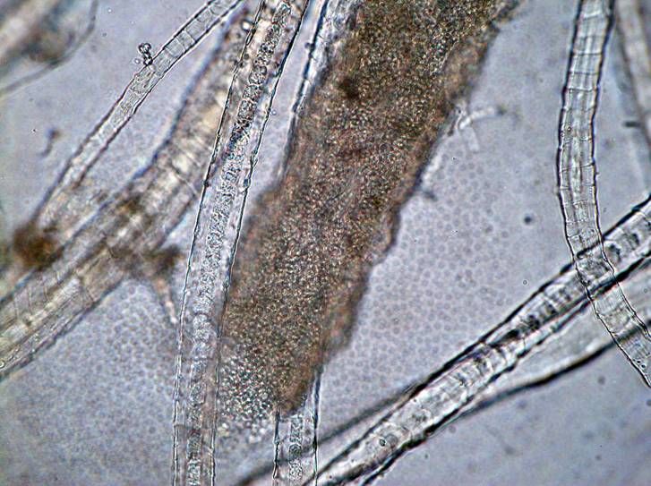
Pathogens of epidermophytosis

Pathogens epidermophytia are dermatophytes, or dermatomycetes. They cause trichophytosis, microsporia, favus and other lesions of the skin, nails and hair. Dermatophytes are divided into three genera: Microsporum, Trichophyton, Epidermophyton, whose representatives differ in the ways of sporulation.

Morphology and physiology of dermatophytes
Dermatophytes have a septic mycelium with arthroconidia, macro- and microconidia. In fungi of the genus Epidermophyton, there are many smooth club-like macroconidia, and in representatives of the genus Microsporum - thick-walled, multicellular spindle-shaped spiked microconidia. For fungi of the genus Trichophyton, large smooth septate macroconidia are characteristic, the fungi reproduce asexually (anamorphs) or sex (teleomorph) pathways. Grow on the environment of Saburo and others. Colonies (depending on the species) are multi-colored, mealy, granular, furry.
Resistance of dermatophytes
Fungi are resistant to drying and freezing. Trichophytons persist in the hair up to 4-7 years. Dermatophytes perish at 100 ° C in 10-20 minutes. Sensitive to the action of UV rays, solutions of alkali, formaldehyde, iodine.
Pathogenesis and symptoms of epidermophytosis
Pathogens live on keratinized substrates (keratophilic fungi). The development of the disease contributes to minor skin damage, maceration, weakened immunity, increased sweating, endocrine disorders and prolonged use of antibiotics. Dermatophytes do not penetrate the basement membrane of the epidermis. In varying degrees, the skin, hair and zeros are affected. There are dermatomycosis of the trunk, limbs, face, feet, brushes, perineum, areas of the beard, scalp, nails (onychomycosis).
Hair struck by mushrooms break off; develop focal alopecia, baldness. The skin is peeling, vesicles, pustules, cracks appear. Itching develops lesions. Inflammation is absent or may be in severe form. With fungal infections of the nails (onychomycosis), the color, transparency, thickness, surface, strength and integrity of the nail plate change. The causative agent of onychomycosis can be any. The development of mycosis is promoted by a decrease in immunity. In humans infected with fungi, IgM-, IgG-antibodies appear, HRT develops.
Epidemiology of epidermophytosis
Pathogens are transmitted by contact with a sick person or animal or by contact with various environmental objects. Infection is possible through household items (combs, towels), as well as in baths, showers and pools.
Anthropophilic dermatophytes are transmitted from person to person. Zoophilic dermatophytes are transmitted to humans from animals. Trichophyton verrucosum is transmitted from cattle (veal deprive). Geofilnye dermatophytes (pathogens microsporia) live in the soil and are transmitted by contact with it.
Microbiological diagnosis of epidermophytosis
Microscopic scrapings from the affected skin, scales, nail plates, hair, treated for 10-15 minutes with 10-15% KOH solution. Preparations are stained with hematoxylin and eosin. You can apply RIF using fluorescent antibodies. With microscopy, the threads of mycelium, arthroconidia, macro- and microconidia, blastospores are revealed. Arthroconidia of the genus Trichophyton can be located in parallel chains outside the hair (ectotriks) and inside the hair (enolizriks). Arthroconidia of the genus Microspomm are arranged mosaically outside the hair. When favus inside the hair, fungal elements and gas bubbles are found.
Sowing is done on nutrient media - Saburo, etc. Growth of fungi is studied after 1-3 weeks of cultivation at 25 ° C. Determine antibodies in the blood serum with the help of RSK, RIGA, RP, RIF, ELISA. They put dermal allergic tests with allergens from mushrooms. A biological sample is placed on laboratory animals (guinea pigs, mice, etc.), infecting them in the skin, hair and nails.
Treatment of epidermophytosis
With dermatophytic fibrous, part of the head is used fluconazole; with nail dermatophyte systemic and local antifungal therapy; When the feet are dermatophytically used antifungal creams and ointments in combination with indications with systemic therapy and antihistamines.
Last reviewed: 01.06.2018
Medical expert editor
Portnov Alexey Alexandrovich
Education: Kiev National Medical University. A.A. Bogomolets, Specialty - "General Medicine"
Other articles on the topic
Malassezia furfur is a kind of fungus that causes dandruff. Fungus can cause seborrhea and atopic dermatitis.


