Medical expert of the article
New publications
Skin: structure, vessels and nerves
Last reviewed: 04.07.2025

All iLive content is medically reviewed or fact checked to ensure as much factual accuracy as possible.
We have strict sourcing guidelines and only link to reputable media sites, academic research institutions and, whenever possible, medically peer reviewed studies. Note that the numbers in parentheses ([1], [2], etc.) are clickable links to these studies.
If you feel that any of our content is inaccurate, out-of-date, or otherwise questionable, please select it and press Ctrl + Enter.
The skin (cutis), which forms the general covering of the human body (integumentum commune), directly contacts the external environment and performs a number of functions. It protects the body from external influences, including mechanical ones, participates in the body's thermoregulation and metabolic processes, secretes sweat and sebum, performs a respiratory function, and contains energy reserves (subcutaneous fat). The skin, which occupies an area of 1.5-2.0 m2 depending on the size of the body, is a huge field for various types of sensitivity: tactile, pain, temperature. The thickness of the skin in different parts of the body is different - from 0.5 to 5 mm. The skin is divided into a superficial layer - the epidermis, formed from the ectoderm, and a deep layer - the dermis (the skin itself) of mesodermal origin.
The epidermis is a multilayered epithelium, the outer layer of which gradually peels off. The epidermis is renewed by its deep germ layer. The thickness of the epidermis varies. On the hips, shoulder, chest, neck and face it is thin (0.02-0.05 mm), on the palms and soles, which experience significant physical stress, it is 0.5-2.4 mm.
The epidermis consists of many layers of cells, united into five main layers: horny, shiny, granular, spiny and basal. The superficial horny layer consists of a large number of horny scales formed as a result of keratinization of the cells of the underlying layers. Horny scales contain the protein keratin and air bubbles. This layer is dense, elastic, does not allow water, microorganisms, etc. to pass through. Horny scales gradually peel off and are replaced by new ones, which approach the surface from the deeper layers.
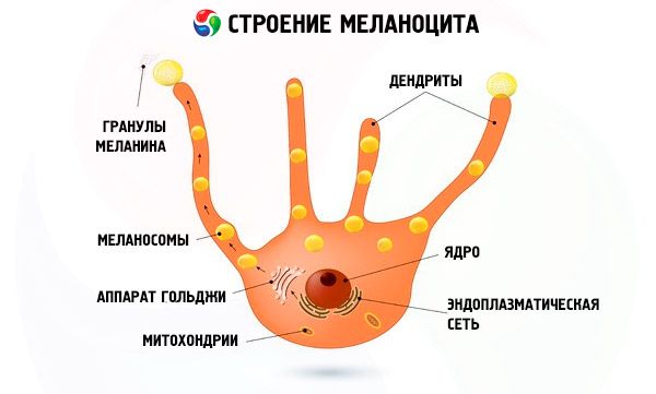
Under the stratum corneum is the stratum lucidum, formed by 3-4 layers of flat cells that have lost their nuclei. The cytoplasm of these cells is impregnated with the protein eleidin, which refracts light well. Under the stratum lucidum is the stratum granulosum, consisting of several layers of flattened cells. These cells contain large grains of keratohyalin, which turns into keratin as the cells move toward the surface of the epithelium. In the depths of the epithelial layer are the cells of the spinous and basal layers, which are united under the name of the germinal layer. Among the cells of the basal layer are pigment epithelial cells containing the pigment melanin, the amount of which determines the color of the skin. Melanin protects the skin from the effects of ultraviolet rays. In some areas of the body, pigmentation is especially well expressed (the areola of the mammary gland, the scrotum, around the anus).

The dermis, or skin proper (dermis, s. corium), consists of connective tissue with some elastic fibers and smooth muscle cells. On the forearm, the thickness of the dermis does not exceed 1 mm (in women) and 1.5 mm (in men), in some places it reaches 2.5 mm (back skin in men). The skin proper is divided into a superficial papillary layer (stratum papillare) and a deeper reticular layer (stratum reticulare). The papillary layer is located directly under the epidermis, consists of loose fibrous unformed connective tissue and forms protrusions - papillae, containing loops of blood and lymphatic capillaries, nerve fibers. In accordance with the location of the papillae on the surface of the epidermis, skin ridges (cristae cutis) are visible, and between them are oblong depressions - skin grooves (sulci cutis). The ridges and grooves are best expressed on the soles and palms, where they form a complex individual pattern. This is used in forensic science and forensic medicine to establish identity (dactyloscopy). In the papillary layer, there are bundles of smooth muscle cells associated with hair follicles, and in some places such bundles lie independently (skin of the face, nipple of the mammary gland, scrotum).
The reticular layer consists of dense, irregular connective tissue containing bundles of collagen and elastic fibers, and a small amount of reticular fibers. This layer passes without a sharp boundary into the subcutaneous base, or cellular tissue (tela subcutanea), containing fat deposits (panniculi adiposi) to a greater or lesser extent. The thickness of the fat deposits is not the same in all places. In the forehead and nose area, the fat layer is weakly expressed, and it is absent on the eyelids and skin of the scrotum. On the buttocks and soles, the fat layer is especially well developed. Here it performs a mechanical function, being an elastic lining. In women, the fat layer is better developed than in men. The degree of fat deposition depends on the type of build and nutrition. Fat deposits (fatty tissue) are a good heat insulator.
Skin color depends on the presence of pigment, which is present in the cells of the basal layer of the epidermis and is also found in the dermis.
Vessels and nerves of the skin
Branches from superficial (cutaneous) and muscular arteries penetrate the skin, which form a deep dermal and superficial subpapillary arterial network in the thickness of the skin. The deep dermal network is located on the border of the skin proper and the subcutaneous fat base. Thin arteries extending from it branch out and supply blood to the fat lobules, the skin proper (dermis), sweat glands, hair, and also form an arterial network at the base of the papillae.
This network supplies blood to the papillae, into which the capillaries penetrate, forming intrapapillary capillary loops that reach the tops of the papillae. From the superficial network, thin vessels branch off to the sebaceous glands and hair roots. Venous blood from the capillaries flows into the veins that form the superficial subpapillary and then the deep subpapillary venous plexus. From the deep subpapillary plexus, venous blood flows into the deep dermal venous plexus and then into the subcutaneous venous plexus.
The lymphatic capillaries of the skin form a superficial network in the reticular layer of the dermis, where the capillaries located in the papillae flow, and a deep network - at the border with the subcutaneous fat tissue. The lymphatic vessels formed from the deep network, connecting with the vessels of the muscle fascia, are directed to the regional lymph nodes.
The skin is innervated by both branches of somatic sensory nerves (cranial, spinal) and fibers of the autonomic (autonomous) nervous system. In the epidermis, papillary and reticular layers there are numerous nerve endings of different structures that perceive touch (touch), pressure, pain, temperature (cold, heat). Nerve endings in the skin are unevenly distributed. They are especially numerous in the skin of the face, palms and fingers, and external genitalia. Innervation of glands, hair-raising muscles, blood and lymphatic vessels is carried out by postganglionic sympathetic fibers that enter the skin as part of the somatic nerves, as well as together with blood vessels. Nerve fibers form plexuses in the subcutaneous fat and in the papillary layer of the dermis, as well as around the glands and hair roots.

