Medical expert of the article

New publications
Rubrophytosis of the skin of feet, hands, face, nails
Last reviewed: 04.07.2025

All iLive content is medically reviewed or fact checked to ensure as much factual accuracy as possible.
We have strict sourcing guidelines and only link to reputable media sites, academic research institutions and, whenever possible, medically peer reviewed studies. Note that the numbers in parentheses ([1], [2], etc.) are clickable links to these studies.
If you feel that any of our content is inaccurate, out-of-date, or otherwise questionable, please select it and press Ctrl + Enter.
Rubrofitia (synonym: rubromycosis) is the most common fungal disease that affects smooth skin, toenails, hands, and vellus hair.
Causes rubrophytes
The causative agent of the disease is the fungus Trichophyton rubrum. This infection accounts for 80-90% of all pathogens causing foot mycosis. Infection occurs in the same way as with athlete's foot (see athlete's foot).
Symptoms rubrophytes
The following forms of rubromycosis are distinguished: rubromycosis of the feet, rubromycosis of the feet and hands, generalized rubromycosis.
Onychomycosis of the feet
Rubromycosis of the feet is the most common. The clinical picture of the disease begins with the lesion of the interdigital folds of the feet. Gradually, the process spreads to the skin of the soles and nail plates (onychomycosis).
The skin of the affected soles is stagnantly hyperemic, moderately lichenified, the skin pattern is enhanced, the surface is usually dry; in the grooves, mucous peeling or peeling in the form of small rings and figures of scalloped outlines is quite well expressed. Over time, the skin pathological process moves to the lateral and dorsal surfaces of the feet. Subjectively, itching of the skin is noted, sometimes excruciating.
The pathological process usually also involves the toenails.
There are three types of nail plate damage: normotrophic, hypertrophic and atrophic.
In the normotrophic type, the nail plate is affected from the lateral (or free) edges in the form of white or yellowish stripes or the same stripes visible in the thickness of the nail plate.
In the hypertrophic type, the nail plate thickens due to subungual hyperkeratosis. It is dull, crumbles from the free edge. The mentioned stripes are also visible in its thickness.
In the atrophic type, most of the nail plate is destroyed, remaining only partially at the nail fold. Sometimes, the nail plate can separate from the nail bed by the onycholysis type.
Rubromycosis of the feet and hands
This form of rubromycosis occurs in patients suffering from mycosis of the feet.
The clinical picture of rubromycosis on the hands is very similar to the manifestation of rubromycosis of the feet. The skin-pathological process is much less pronounced due to repeated hand washing during the day. The presence of foci draws attention: foci with an intermittent inflammatory ridge along the periphery and on the back of the hand, a reddish-bluish background of the skin of the palms. On the surface of the elements, mucous peeling is noted in varying degrees of severity. When the nail plates of the hands are involved in the pathological process, they are also affected by the normotrophic, hypertrophic or atrophic type.
Rubromycosis generalized
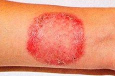
Generalization of fungal infection is observed in patients suffering from rubromycosis of the skin of the feet or onychomycosis for a long time. The spread of rubromycosis is facilitated by pathology of internal organs, endocrine system, insufficiency of the immune system. Large folds are most often affected, especially the inguinal-femoral, buttocks and shins, but foci can also be found on other areas of the skin. At first, pink or pink-red spots of rounded outlines with a bluish tint appear, clearly delimited from healthy skin. Later, the color of the foci becomes yellowish-red or brown. They are slightly infiltrated, their surface is covered with small scales, and along the periphery there is an intermittent scalloped ridge consisting of small papules, vesicles and crusts. As a result of peripheral growth and fusion with each other, the spots occupy large areas. Deep lesions of the red trichophyton, mainly of the shins, buttocks and forearms, are considered a follicular-nodular variety of the disease. The rash is accompanied by significant itching, the process is prone to relapse, especially in the warm season. In the generalized form, vellus hair is affected. It loses its shine, becomes dull, breaks off (sometimes in the form of "black dots").
Of great importance in diagnosing the disease are the detection of the fungus during microscopic examination of pathological material (scales, vellus hair) and sowing the material on a nutrient medium to obtain a culture of red trichophyton.
In most patients, the manifestations of generalized rubromycosis develop after the presence of lesions of the skin and nails of the feet (or feet and hands) for a more or less long time (from several months to 5-10 years or more) against the background of pathology of internal organs, the endocrine and nervous system, trophic skin disorders or due to other changes in the body. For example, the development of generalized manifestations of rubromycosis is often facilitated by long-term treatment with antibiotics, cytostatic and steroid drugs.
Trichophyton rubra causes both superficial and deep lesions of smooth skin, which is sometimes observed in the same patient. Thus, rashes in the inguinal and intergluteal folds and deep (nodular-nodular) lesions on the shins or other areas of the skin may occur simultaneously.
Deep lesions of the red grichophyton mainly of the shins, buttocks and forearms are considered as a follicular-nodular variety of the disease. In this form, along with papular-follicular elements, there are also deeper elements that tend to group, are located in the form of arcs, open tracks and garlands. The rash is accompanied by significant itching. The process tends to relapse, especially in the warm season. Foci of this form of rubromycosis can simulate indurative erythema of Bazin, nodular erythema, papulonecrotic tuberculosis (often cicatricial changes remain at the site of the foci), nodular vasculitis, deep pyoderma, leukemids and manifestations of other dermatoses. For example, when rubromycosis is localized on the skin of the face, the lesions can be very reminiscent of lupus erythematosus, tuberculous lupus, manifestations of staphylococcal sycosis, and even pigment xeroderma in the elderly.
Generalized rubromycosis can certainly occur without the formation of deep foci. In such cases, the lesions in clinical manifestations can be very close to eczema, neurodermatitis, parapsoriasis, psoriasis, annular granuloma, Devergie's lichen pilaris, etc. Exudative manifestations of rubromycosis can also be observed - small vesicular rashes and crusts on the feet, hands and other areas of the skin.
It should be noted that with exudative manifestations of rubromycosis, a number of patients may develop secondary (allergic) rashes on the skin of the trunk and extremities that do not contain fungal elements.
The most common forms of rubromycosis are those where the lesions are deep red (often with a bluish tint), merging with each other, and have more or less pronounced peeling on the surface. Clinical varieties of the disease include mycotic erythroderma and palmar-plantar-inguinal-gluteal syndrome. This syndrome, which is observed in many patients with generalized rubromycosis, usually affects the skin of the feet, palms, and nail plates.
Lesions of large folds - intergluteal, inguinal-femoral, skin of the buttocks, under the mammary glands usually occur after a more or less long existence of foci of mycosis on the feet and palms. The foci seem to emanate from the depths of large folds, spreading to the inner quadrants of the buttocks and then to the outer ones. The surface of the foci is yellowish-red or brown. They are slightly infiltrated, slightly flaky. The edges of the foci are slightly elevated, having an intermittent scalloped ridge consisting of small papules and crusts. Usually the ridge has a more intense reddish-bluish tint than the lesion itself.
Diagnostics rubrophytes
Of great importance in diagnosing the disease is the detection of the fungus during microscopic examination of pathological material (scales, vellus hair) and sowing the material on a nutrient medium to obtain a culture of red trichophytope.
The diagnosis of rubromycosis of the feet (or feet and hands) is based on a fairly characteristic clinical picture and the detection of fungal elements in the foci. But often, especially in the case of latent or atypically occurring rubromycosis, the result of cultural studies is decisive for making a diagnosis. These studies are especially important in dyshidrotic forms of rubromycosis, which are very similar (if not clinically identical to it) to epidermophytosis of the feet caused by Trichophyton interdigitale.
What do need to examine?
How to examine?
Differential diagnosis
When conducting a differential diagnosis of rubromycosis, it is necessary to keep in mind superficial (anthropophilic) trichophytosis, as well as limited forms of infiltrative-suppurative (zoophilic) trichophytosis. It should also be remembered that the rather rarely observed lesions of the scalp in rubromycosis may resemble foci of microsporia.
The differential diagnosis of rubromycosis of the feet (or feet and hands) should first be carried out with epidermophytosis of the feet (and epidermophytids), trichophytosis caused by fungi of the anthropophilic group, palmar-plantar hyperkeratosis, psoriasis and eczema of this localization.
It should be borne in mind that lesions of the interdigital folds and nail plates can be caused by yeast-like fungi of the genus Candida, mold fungi, and other dermatophytes.
 [
[ Who to contact?
Treatment rubrophytes
Treatment of athlete's foot and rubrofitia should be etiotropic, pathogenetic and symptomatic. Treatment should begin with external therapy. In acute inflammatory processes with oozing, lotions of 2% resorcinol, boric acid, 0.25% silver nitrate are prescribed. The cover of the vesicles (blisters) is pierced with a needle or cut off with scissors, observing aseptic rules. Then, solutions of aniline dyes are used (Costellani paint, methylene blue, brilliant green, etc.). For etiotropic treatment, creams and ointments containing antimycotics are prescribed (1% cream or derm-gel of lamisil, travogen, zalain, etc.). In the presence of severe inflammation and the addition of a secondary infection, ointments or creams containing corticosteroids and antibiotics are prescribed together with antimycotics (travocort, gentriderm, triderm, etc.). In order to dry out foci of weeping, an antifungal drug is widely used - nitrofungin-neo in the form of a solution and spray. Lamisil is used in the form of derm-gel or 1% cream once a day for 7 days. When using mix forms of lamisil, by the end of therapy in patients with foot mycosis, clinical recovery occurred in 82%, mycological - in 90% of patients. By the end of the second week, clinical and mycological recovery was noted in all patients. According to many scientists, such a pronounced effect is due to the lipophilic and keratophilic properties of the drug, rapid penetration and long-term preservation of a high concentration of terbinafine in keratinized skin. Lamisil can be used for mycosis of the feet complicated by a secondary infection, since it has been proven that the drug has anti-inflammatory activity like cycloripoxolamine and an antibacterial effect like 0.1% gentamicip cream.
In the erythematous-squamous form of mycosis of the feet, accompanied by cracks, the use of Lamisil in the form of a 1% cream for 28 days contributes not only to clinical and mycological cure, but also to the healing of superficial and deep cracks. Therefore, Lamisil, in addition to antifungal, antibacterial and anti-inflammatory properties, has the ability to stimulate regenerative processes in the skin.
Systematic symptomatic treatment includes the use of desensitizing, antihistamine, sedative agents and vitamins, since the causative agents of this fungal infection have pronounced antigenic properties.
If there is no effect from external agents, you should switch to taking systemic antimycotics.
Currently, the following systemic antimycotics are used as etiotropic agents: terbinofine (Lamisil), itraconazole (Tecnazole, Orungal), griseofulovin, etc.
Lamisil for athlete's foot without nail plate damage is prescribed in a daily dose of 250 mg for 14 days. For mycosis of the feet, itraconazole (teknazole, orungal) is used at 100 mg once a day for 15 days.
For foot onychomycosis, Lamisil is prescribed at 250 mg per day for 3 months, and for hand onychomycosis - for 1.5 months. Itracopazole (Teknazole, Orungal) is used at 200 mg 2 times per day for a week (one course), then a 3-week break is taken. For foot onychomycosis, 3 courses of treatment are prescribed, and for hand onychomycosis - 2 courses.
Considering the pronounced allergenic properties of the pathogen, it is necessary to prescribe (especially in the presence of mycids) desensitizing agents and antihistamines, sedatives, B vitamins, rutin, ascorbic acid. In the case of secondary pyogenic infection, short-term courses of broad-spectrum antibiotics are indicated.
It is necessary to eliminate concomitant diseases (diabetes mellitus, endocrine, immune disorders, impaired microcirculation of the lower extremities, etc.).
General prevention requires hygienic maintenance and regular disinfection of baths (floors, rugs, wooden grates and pads, benches, basins), showers and swimming pools, medical examinations of the personnel serving them, timely treatment and medical examination of patients. Personal prevention consists of using only your own shoes, observing the rules of personal hygiene of the skin of the feet, and disinfecting shoes. Wipe the insole and lining of the shoe with a cotton swab soaked in 25% formalin solution or 0.5% chlorhexidine bigluconate solution. Then place the shoes in a polyethylene bag for 2 hours and air until dry. Disinfect socks and stockings by boiling for 10 minutes. To prevent relapses of epidermophytosis, after the symptoms of the disease have disappeared, lubricate the skin of the feet with antimycotic agents for 2-3 weeks. For prevention purposes, nitro-fungin-neo is widely used in the form of a solution or spray.