Medical expert of the article

New publications
Respiratory (lung) examination
Last reviewed: 06.07.2025

All iLive content is medically reviewed or fact checked to ensure as much factual accuracy as possible.
We have strict sourcing guidelines and only link to reputable media sites, academic research institutions and, whenever possible, medically peer reviewed studies. Note that the numbers in parentheses ([1], [2], etc.) are clickable links to these studies.
If you feel that any of our content is inaccurate, out-of-date, or otherwise questionable, please select it and press Ctrl + Enter.
The doctor receives certain objective information already during the conversation with the patient and the general examination: the general appearance of the patient, position (active, passive, forced on the sore side in pleurisy and pleuropneumonia), the condition of the skin and visible mucous membranes (cyanosis, pallor, the presence of herpetic eruptions on the lips, wings of the nose and unilateral hyperemia of the face as signs accompanying pneumonia). Particular attention is paid to the change in the shape of the nails like watch glasses and the terminal phalanges of the fingers like drumsticks (Hippocratic fingers), characteristic of chronic pulmonary suppurations (bronchiectasis, lung abscess), as well as bronchogenic cancer, fibrosing alveolitis.
This symptom (especially in relation to bronchogenic carcinoma) is also called pulmonary hypertrophic osteoarthropathy (meaning the possibility of damage to other bones with pain in them). However, it should be remembered that this symptom may also be associated with non-pulmonary diseases (blue congenital heart defects, subacute infective endocarditis, liver cirrhosis, nonspecific ulcerative colitis, subclavian artery aneurysm, chronic hypoxia in high altitude conditions). There may be familial cases of such changes.
In some lung diseases, eye lesions are found: nonspecific keratoconjunctivitis in primary tuberculosis, iridocyclitis also in tuberculosis and sarcoidosis.
Examination of the lymph nodes is important: enlargement of the supraclavicular lymph nodes can be observed in lung tumors (metastases), lymphoma, sarcoidosis, tuberculosis and requires a biopsy.
Some skin changes allow us to suspect or help us understand the development of a pulmonary process. Thus, erythema nodosum is a very characteristic non-specific sign of sarcoidosis (as well as peculiar specific sarcoid nodules); in bronchogenic cancer, metastatic nodules can be detected in the skin; lung damage in systemic diseases is accompanied by the appearance of various rashes on the skin ( hemorrhagic vasculitis, etc.).
Diseases associated with Hippocratic fingers syndrome
Respiratory diseases:
- Bronchogenic cancer.
- Chronic suppuration ( bronchiectasis, abscess, empyema ).
- Fibrosing alveolitis.
- Asbestosis.
Cardiovascular diseases:
- Congenital heart defects (blue type).
- Subacute infective endocarditis.
- Subclavian artery aneurysm.
Diseases of the gastrointestinal tract:
- Cirrhosis.
- Non-specific ulcerative colitis.
- Malabsorption syndrome (steatorrhea).
- Familial (congenital) changes in phalanges. High altitude hypoxia.
During a general examination, important signs such as cyanosis and edema are revealed.
Cyanosis (blueness) is a peculiar change in skin color, usually best detected on the lips, tongue, ears, nails, although sometimes it is total. Pulmonary cyanosis most often occurs with alveolar hypoventilation or imbalance between ventilation and perfusion. The severity of cyanosis depends on the content of reduced hemoglobin in the tissue capillaries, so patients with anemia do not look cyanotic even with low PO2, and vice versa, with polycythemia, cyanosis is usually easily detected, although the oxygen tension in the blood is normal or even increased. Local cyanosis of the extremities may be associated with shunting of a large part of the blood that does not reach the extremities (shock).
For lung diseases (primarily obstructive), as well as pneumonia, bronchial asthma, fibrosing alveolitis, so-called central cyanosis is characteristic, developing in connection with peripheral vasodilation and accumulation of carbon dioxide in the blood. Peripheral cyanosis with a predominant change in the color of the face, neck, and sometimes the upper limbs is most often caused by compression of the superior vena cava. Such compression (for example, in lung cancer ) is accompanied by local edema and the development of venous collaterals on the anterior surface of the chest.
Usually, edema syndrome in a patient with lung disease is a sign of right ventricular heart failure.
The examination of the respiratory system begins with a question about nasal breathing, the presence of nosebleeds. At the same time, the voice and its changes, in particular hoarseness, are assessed.
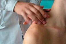
The doctor obtains important data from examination and palpation of the chest, percussion and auscultation of the lungs.
The chest is examined during normal breathing and under conditions of increased breathing. The depth and frequency of breathing are determined (normally the number of respiratory movements and the pulse rate are related as 1:4), the degree of acceleration, the ratio of inhalation and exhalation time (exhalation is prolonged during obstruction of small bronchi; difficulty in inhalation up to whistling, noisy, so-called stridor breathing during narrowing of the trachea and large bronchi), symmetry and the nature of respiratory movements of the chest.
It should be remembered that during breathing, the intrathoracic pressure constantly changes, due to which air enters and leaves the pulmonary alveoli through the respiratory tract. When inhaling, the diaphragm descends, the chest moves up and to the sides, which increases the intrathoracic volume, reduces the intrathoracic pressure, and air enters the alveoli. Under normal conditions, the supply of the required amount of oxygen and the removal of carbon dioxide is ensured by a minute breathing volume of 5-6 liters of air.
An increase in minute ventilation is primarily achieved by rapid breathing (tachypnea), but without increasing its depth, which, for example, occurs in widespread pulmonary fibrosis, pleural diseases, chest rigidity, pulmonary edema. Breathing becomes more frequent (tachypnea) and deeper (hyperpnea) - the so-called "air hunger" or Kussmaul breathing, for example, in diabetic ketoacidosis, renal metabolic acidosis. Minute ventilation changes in diseases of the central nervous system: in meningitis it increases, in tumors and hemorrhage due to increased intracranial pressure it decreases. Ventilation inhibition is observed under the influence of anesthetics and other drugs.
During examination, forced exhalation can be detected - an effort required to increase intrathoracic pressure in order to overcome the resistance to the free flow of air outward, which is typical for chronic obstructive pulmonary diseases ( chronic bronchitis, pulmonary emphysema, bronchial asthma). In this case, in addition to lengthening the exhalation, the inclusion of auxiliary muscles of the neck, shoulder girdle, and intercostal spaces is revealed.
The shape of the chest, its mobility during breathing (participation in the act of breathing) are assessed. Normosthenic, asthenic and hypersthenic chests are distinguished, which correspond to other signs of a certain constitutional type of a person. Thus, due to the proportional relationship of the anterior-posterior and transverse sizes in the normosthenic form, the epigastric angle formed by the costal arches is 90°, the ribs have an oblique direction, the supra- and subclavian fossae are moderately expressed, the shoulder blades are tightly adjacent to the back. In contrast, in the asthenic form, the chest is flat, the epigastric angle is less than 90°, the ribs are located more vertically, the shoulder blades have the appearance of wings, and in the hypersthenic form, these landmarks have the opposite direction.
Depending on the damage to the lungs and pleura or changes in the skeletal system, the above-mentioned types of chest may acquire specific pathological forms. Paralytic (more pronounced signs of the asthenic type) occurs with chronic wrinkling (sclerosing) processes in the lungs or pleura that usually begin in childhood; barrel-shaped, emphysematous (sharply pronounced signs of the hypersthenic type) develops as a result of widespread hyperaerotic expansion (emphysema) of the lungs, caused by the loss of elasticity of the lung tissue and the inability of the lungs to collapse on exhalation, which is accompanied by a decrease in respiratory excursion characteristic of an emphysematous chest. Incorrect formation of the skeleton in rickets in childhood leads to the so-called rachitic chest with a protruding sternum ("chicken breast"). In connection with changes in the skeletal system, a distinction is made between a funnel-shaped chest (inward depression of the sternum - "shoemaker's chest") and a scaphoid (a common boat-shaped depression of the upper part of the chest wall from the front). Of particular importance are changes in the shape of the chest in connection with curvatures of the thoracic spine: lordosis (a forward convexity of the spine), kyphosis (a backward convexity of the spine), scoliosis (a sideways curvature of the spine), but especially kyphoscoliosis, when the heart and large vessels, including the pulmonary vessels, find themselves in unusual conditions, which leads to the gradual development of right ventricular heart failure ("kyphoscoliotic heart").
Examination, especially dynamic, using deep breathing, allows to reveal asymmetries of the chest: asymmetry of shape (bulging, retraction) and asymmetry of participation in the act of breathing. Bulging of the corresponding half of the chest wall with smoothing of the intercostal spaces usually develops in the presence of fluid ( pleurisy, hydrothorax) or gas ( pneumothorax ) in the pleural cavity, sometimes with a widespread infiltrate (pneumonia) or a large lung tumor. Retraction of one half of the chest is observed with a widespread fibrous process that shrinks the lung and the development of obstructive atelectasis (collapse) of the lobe of the lung due to blockage of the bronchus draining this lobe (endobronchial tumor, external compression, foreign body in the lumen of the bronchus). Usually in all these cases the half of the chest corresponding to the deformation lags behind in breathing or does not participate in the act of breathing at all, and thus the detection of this phenomenon has important diagnostic significance.
 [
[