Medical expert of the article
New publications
Cervical hernia
Last reviewed: 29.06.2025

All iLive content is medically reviewed or fact checked to ensure as much factual accuracy as possible.
We have strict sourcing guidelines and only link to reputable media sites, academic research institutions and, whenever possible, medically peer reviewed studies. Note that the numbers in parentheses ([1], [2], etc.) are clickable links to these studies.
If you feel that any of our content is inaccurate, out-of-date, or otherwise questionable, please select it and press Ctrl + Enter.

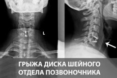
Cervical herniation involves displacement of the pulposus (gelatinous) nucleus of the intervertebral disc beyond the surrounding fibrous ring.
What are the dangers of cervical herniated discs? The protrusion of part or all of the nucleus pulposus through the fibrous ring of the intervertebral disc can lead to nerve compression or direct compression of the spinal cord located in the spinal canal. In addition, when a herniated cervical vertebra puts pressure on one of the vertebral arteries, the cerebral circulation may be impaired.
Causes of the cervical hernias
Many vertebrologists consider age to be the main cause of herniated disc of the cervical spine, because over time - in the course of natural aging or wear and tear - degenerative and dystrophic changes occur in the discs: they gradually lose fluid volume (the pulp nuclei, located in the center of the discs, is almost two-thirds composed of chondroitin-sulfate bound water). [3]
Part of the negative changes in the intervertebral disc, which cause its weakening and bulging of the pulposus nucleus, is due to changes in the composition of collagen, the main structural protein of the extracellular matrix of various connective tissues. The connection of herniation with the decrease of type II collagen - the main component of cartilage extracellular matrix cross-linked with proteoglycans (sulfated glycosaminoglycans) and the increase of type I collagen, which has a larger diameter of fibrils and a different system of their arrangement and is found in the whole organism, except for cartilage tissue. With age, the synthesis of type II fibrillar collagen by chondrocytes (cartilage tissue cells) decreases, which is obviously associated with a decrease in the amount of mRNA (matrix ribonucleic acid) of type II procollagen.
In addition, the causes of intervertebral disc degeneration can be genetically determined. These are type II collagenopathies with a mutation in the COL2A1 gene, which encodes the protein filaments (alpha chains) that make up type II collagen.
Matrix metalloproteinase (MMP) expression may also be increased due to mutations in a group of genes encoding proteins of this proteolytic enzyme. It participates in normal physiological processes of tissue remodeling, but with increased activity it destroys collagen and proteoglycans, which negatively affects the condition of intervertebral discs.
Intervertebral herniation of this localization is etiologically often associated with trauma to the cervical spine, as well as osteochondrosis of the cervical vertebrae. [4]
Risk factors
Factors that increase the risk of a cervical herniated disc include:
- Age 50+;
- Having a family history of vertebral herniation;
- Curvature of the spine - scoliosis in the cervical vertebrae;
- Excessive external influences on the cervical region (static load, whole body vibration, repetitive movements, occupational movements and positioning of the head and neck);
- Autoimmune diseases, primarily systemic lupus erythematosus and rheumatoid arthritis;
- Sedentary lifestyle;
- Deficiency of vitamin C (cofactor of collagen synthesis by chondrocytes).
Pathogenesis
The cervical spinal column has seven cervical vertebrae (C1-C7); like all vertebrae, they are separated from each other by fibrous-cartilaginous intervertebral (intervertebral) discs, which serve a shock-absorbing function and provide the vertebrae with relative mobility.
Intervertebral discs have an outer fibrous ring composed of connective tissue cells, and a pulposus nucleus, the inner gel-like portion of the disc, which is composed of water, type II collagen, chondrocyte-like cells, and proteoglycans, particularly aggrecan. This glycosaminoglycan contains multiple chains of negatively charged chondroitin sulfate and keratansulfate that bind water and thereby hold together a network of collagen fibrillar fibers. This composition provides the nucleus pulposus with elasticity, flexibility under load and resistance to compression - redistributing the load to the annulus fibrosus and cartilaginous closure plates that attach the intervertebral discs to neighboring vertebrae. [5]
Aging modifies collagen fibrils with accumulation of non-enzymatic glycation end products that increase the stiffness of collagen fibers.
The pathogenesis of degenerative and dystrophic changes in the structures of the intervertebral disc - the nucleus pulposus and the annulus fibrosus - is usually associated with the loss of proteoglycan molecules that bind water. The loss of water causes the nucleus to become fibrous and stiffer, which reduces its ability to bear stress, and the excess load is transferred to the fibrous ring. But the degenerative process also affects the structure of the fibrous ring, in the form of its thinning, loss of elasticity and the formation of microcracks, to which the pulposus nucleus is displaced. There is disc protrusion - its displacement into the spinal canal without rupture of the surrounding fibrous ring. And when the fibrous ring is ruptured, the nucleus is displaced into the epidural space of the spinal canal, where the spinal cord is located. [6]
Herniations are more likely to occur posterolaterally, where the fibrous ring is thinner and not supported by the longitudinal ligament on the posterior surface of the vertebral bodies.
Symptoms of the cervical hernias
Herniated discs are often asymptomatic or may cause symptoms in the form of pain with flexion, extension and rotation of the neck, which may irradiate to the upper extremities. Patients may also experience muscle weakness, numbness and paresthesias (impaired skin sensation) in the upper extremities.
Not only the rupture of the fibrous ring causes pain in cervical herniation. Innervation of the pulp nuclei and intervertebral discs is provided by the sinuvertebral (recurrent spinal) nerves and gray connecting branches of the neighboring paravertebral ganglia of the sympathetic trunk. Therefore, due to irritation of the sensory nerves in the disc, pain occurs, and when the disc compresses or irritates a nerve root, segmental cervical radiculopathy [7] - with pain (dull, aching and difficult to localize or sharp and burning); limitation of neck mobility; weakness and numbness in the neck, shoulders or arms.
There may also be cervical herniated disc headaches and cervical discogenic dizziness.
C3-C4 herniation of the cervical spine can manifest with pain at the base of the neck up to the shoulder bone and in the clavicle area; weakness of the lash muscles of the head and neck, the trapezius and longest muscle of the neck, the scapulae levator muscle, as well as chest pain.
When the pulposus nucleus is displaced into the hole between the vertebrae C4-C5, neck pain radiates to the shoulder, weakness is felt in the deltoid muscle of the shoulder, and impaired sensation touches the outer surface of the shoulder.
Cervical disc herniations most commonly occur between the C5-C6 and C6-C7 vertebral bodies. C5-C6 cervical disc herniation is manifested by headaches, pain in the neck, scapula and arm; weakness of the biceps muscle of the shoulder, numbness of the fingers of the hand (thumb and index finger).
Headaches and cervical pain, which irradiate under the scapula and into the shoulder, and on the dorsal surface of the forearm - to the index and middle fingers of the hand; impaired sensation of the fingers of the hand, weakness of the triceps muscle of the shoulder, stiffness of head movements is manifested by herniation of the cervical spine C6-C7.
Symptomatology depends on the direction of displacement of the pulposus nucleus and the stage of cervical herniation:
- If the displacement of the nucleus pulposus does not exceed 2 mm and the fibrous ring is unchanged, it is stage 1;
- If the inner gel-like part of the disc bulges beyond the fibrous ring by 4 mm, stage 2 is defined;
- At stage 3, the pulp nucleus is displaced by 5-6 mm with rupture of the fibrous ring;
- When the displacement is more than 6 mm, stage 4 hernia is diagnosed.
According to the direction of displacement of the pulposus nucleus, specialists determine the types or kinds of cervical spinal herniations:
- Median cervical herniation: bulge in the center of the spinal canal of the spine (running behind the vertebral bodies) in the direction of its axis;
- Paramedian herniation of the cervical spine (right or left-sided): displacement is observed in the center and on the side of the spinal canal;
- Posterior cervical hernias are defined when the nucleus of the intervertebral disc bulges to the rear;
- Posterolateral (posterolateral) hernias are defined in cases where the pulp nucleus is displaced posteriorly and laterally relative to the spinal axis;
- Dorsal herniation of the cervical spine: the bulge is directed toward the spinal cord canal;
- Far lateral or foraminal herniation of the cervical spine is defined when a disc fragment bulges below and just to the side of the arcuate (facet) joint of the vertebra in the area of the intervertebral (foraminal) hole.
- Diffuse cervical herniation is an irregular bulging of the disc in different directions.
When a fragment separates (sequestration) from a displaced disc nucleus, a sequestered cervical herniation is defined. The opening through which the fragment of the pulp nucleus exits is called the "herniation gate".
Complications and consequences
The main complications of cervical disc herniation of the cervical spine include:
- Segmental radiculopathy (radicular syndrome) with paresthesias, weakness and paralysis of muscles of the neck, upper extremities and facial muscles;
- Compression vertebrogenic myelopathy (which develops due to compression of the spinal cord);
- Anterior spinal or vertebral artery syndrome;
- Thyroid disorder.
Diagnostics of the cervical hernias
In the diagnosis of cervical spine herniation, a detailed patient history and physical examination are important, with emphasis on neurologic examination using provocative tests (Sperling, Hoffman, Lhermitte's symptom).
Instrumental diagnostics - (MRI) magnetic resonance imaging of the cervical region is used to visualize herniated displacement; electromyography and CT myelography may be required. [8]
In addition, patients with alarming symptoms may require laboratory tests: blood tests (total, blood counts and C-reactive protein) as well as MMP (matrix metalloproteinase) tests.
Differential diagnosis
Differential diagnosis is made with osteochondrosis, spondylosis [9] and vertebral spondyloarthrosis; retrolisthesis (dislocation) of the cervical vertebrae, facet syndrome, spinal canal stenosis and cervical foraminal stenosis, myogelosis of the cervical spine, cervical migraine (Barre-Lieu syndrome), neck myositis and syringomyelia of the cervical spinal cord.
Treatment of the cervical hernias
Drug treatment is symptomatic, in which drugs of various pharmacological groups are used. [10]
First of all, painkillers are prescribed for cervical herniation, and these are NSAIDs (non-steroidal anti-inflammatory drugs): ibuprofen, Ketoprofen, Dexketoprofen, neurodiclovit (with diclofenac), meloxicam and others.
Gels and ointments can be used externally for cervical herniated discs: dolgit and Deep Relief (with ibuprofen), Febrofid or ultrafastin (with ketoprofen), naproxen gel, pain relieving ointments vipratox, Viprosal, Apizartron, etc. More information in the article - effective ointments for neck pain.
In cases of intolerable pain, vertebral and paravetrebral blockade for cervical herniation is performed - local anesthetic agents (Novocaine) or corticosteroids (Prednisolone or Hydrocortisone).
If muscle spasms are present, myorelaxants are prescribed, for example, Cyclobenzaprine (Myorix) or tizanidine.
Can chondroprotectors for the spine be used for cervical hernia? Since the results of studies of the effectiveness of the combination of chondroitin sulfate and glucosamine (included in the composition of chondroprotective agents) for hernias are ambiguous, vertebrorologists are in no hurry to prescribe them to patients with vertebral hernias of any localization. The reason is that chondroprotectors (taken internally or administered parenterally) cannot restore intervertebral discs.
Physical therapy treatment for cervical spinal herniation utilizes techniques such as:
- Electrophoresis (with analgesics or corticosteroids) and ultraphonophoresis;
- Magnetic field exposure - magnetotherapy or magnetopuncture;
- Acupuncture or acupuncture;
- Therapeutic massage;
- Hirudotherapy (medical leeches are placed on the neck, which activates the trophism of the periorbital tissues).
Regarding the fact that manual therapy can help with cervical herniation, most vertebrologists express their doubts. And not unreasonably: firstly, mechanical impact on the cervical spine does not eliminate the cause of herniation; secondly, in a significant proportion of patients, manual manipulations only increase neck pain. [11]
LFC for cervical hernia is therapeutic gymnastics, which includes exercises for the long muscles of the neck and head and deep muscles of the neck: smooth turns of the head (right-left) and head tilts (forward-backward).
To reduce the load on the vertebrae, muscles and ligaments of the neck during sleep should be used semi-rigid orthopedic pillow for cervical hernia (with elastic fillers).
A rigid corset for cervical herniation is not recommended to wear, but a cervical bandage can be used in the exacerbation of pain syndrome - to immobilize the vertebrae and reduce the load on them.
Associated with sudden movements, running, jumping and lifting weights, sports for cervical hernia are contraindicated, and experts recommend swimming and walking.
Surgical intervention - cervical herniated disc surgery - is performed only in cases of severe cervical radiculopathy not amenable to conservative treatment. [12], [13]
The following types of operations may apply:
- Laminectomy - surgical removal of a fragment of vertebral bone above the nerve root;
- Discectomy with spondylosis - removal of part or all of the intervertebral disc and fusion of adjacent vertebrae;
- Endoscopic removal of cervical herniation - removal of the displaced part of the pulposus nucleus of the disc.
Also read - spinal Hernia Treatment
Prevention
The spine requires attention, and if you avoid trauma to its cervical region and timely treat cervical osteochondrosis, it is possible to prevent the formation of cervical herniation.
You need to watch your posture and exercise. Since cartilage tissue does not contain blood vessels, nutrients reach the chondrocytes by diffusion, which is facilitated by exercise.
Forecast
Pain, mobility limitation and radiculopathy resulting from a herniated disc usually resolve on their own within six weeks in most patients, aided by enzymatic resorption of the herniated cervical spine, as a result, the herniated bulge may significantly shrink or disappear completely. [14], [15]
However, if symptoms occur for more than a month and a half, the prognosis is less comforting. In severe cases, radicular syndrome or compression of the spinal cord can lead to disability, and disability for cervical herniation is not excluded.
Cervical herniation and the army. In the presence of lesions of the intervertebral discs, the question of suitability, limited suitability or unsuitability for military service is decided by the military medical commission depending on the symptoms present.

