Medical expert of the article

New publications
Cervical vertebrae
Last reviewed: 06.07.2025

All iLive content is medically reviewed or fact checked to ensure as much factual accuracy as possible.
We have strict sourcing guidelines and only link to reputable media sites, academic research institutions and, whenever possible, medically peer reviewed studies. Note that the numbers in parentheses ([1], [2], etc.) are clickable links to these studies.
If you feel that any of our content is inaccurate, out-of-date, or otherwise questionable, please select it and press Ctrl + Enter.
The cervical vertebrae(vertebrae cervicales) experience less stress than the other parts of the spine, so they have a small body. The transverse processes of all cervical vertebrae have a transverse process opening (foramen processus transversus). The process ends in tubercles - anterior and posterior. The anterior tubercle of the sixth cervical vertebra is well developed, it is called the carotid tubercle. If necessary, the carotid artery, passing in front of this tubercle, can be pressed against it. The articular processes of the cervical vertebrae are quite short. The articular surfaces of the upper articular processes are directed backward and upward, and of the lower articular processes - forward and downward. The spinous processes of the cervical vertebrae are short, bifurcated at the end. The spinous process of the seventh cervical vertebra is longer and thicker than that of the adjacent vertebrae. It is easily palpable in humans, which is why it is called the protruding vertebra (vertebra prominens).

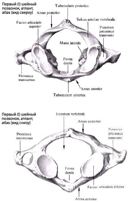
Atlas (atlas) - the 1st cervical vertebra - has no body, since in the embryonic period it fused with the body of the 2nd cervical vertebra, forming its odontoid. The atlas consists of the anterior and posterior arches (arcus anterior et posterior), connected on the sides by two thickenings - lateral masses (massae laterales). The vertebral opening is large and round. On the anterior arch, the anterior tubercle (tuberculum anterior) is located in front. On the inner (posterior) surface of the anterior arch, there is a depression - the pit of the tooth (fovea dentis). It is intended for connection with the odontoid of the 2nd cervical vertebra. On the posterior arch of the atlas, there is a posterior tubercle (tuberculum posterius). It is an underdeveloped spinous process. Above and below, on each lateral mass, there are upper and lower articular surfaces. The upper articular surfaces (facies particulares superiores) are oval and connect with the condyles of the occipital bone. The lower articular surfaces (facies articulates inferiores), on the contrary, are rounded and are intended for articulation with the articular surfaces of the second cervical vertebra. On the upper surface of the posterior arch, the groove of the vertebral artery (sulcus a.vertebralis) is visible on both sides.
Axial - the second cervical vertebra (axis) is distinguished by the presence of a dens - a process extending upward from the body of the vertebra. The dens has an apex and two articular surfaces - anterior and posterior. The anterior articular surface (facies articularis anterior) articulates with the fossa on the posterior surface of the first cervical vertebra, the posterior articular surface (facies articularis posterior) - with the transverse ligament of the atlas. On the sides, the body of the axial vertebra has articular surfaces on top for articulation with the atlas. The lower articular surfaces of the axial vertebra serve for articulation with the third cervical vertebra.
 [
[ Where does it hurt?
What do need to examine?