Medical expert of the article
New publications
Vertebral column (spine)
Last reviewed: 04.07.2025

All iLive content is medically reviewed or fact checked to ensure as much factual accuracy as possible.
We have strict sourcing guidelines and only link to reputable media sites, academic research institutions and, whenever possible, medically peer reviewed studies. Note that the numbers in parentheses ([1], [2], etc.) are clickable links to these studies.
If you feel that any of our content is inaccurate, out-of-date, or otherwise questionable, please select it and press Ctrl + Enter.
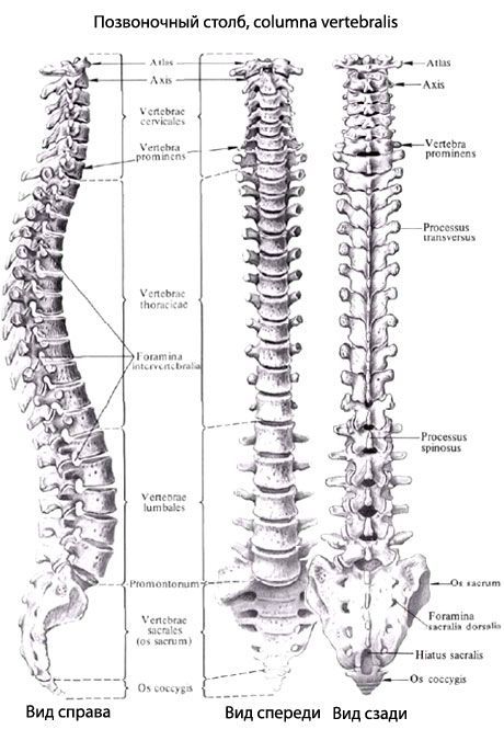
The spine (vertebral column, columna vertebralis) is formed by 33-34 vertebrae, of which 7 are cervical, 12 thoracic, and 5 lumbar.
The most important supporting structure of the human body is the spine. Without it, the average person would have no chance of walking or running, or even standing without assistance.
In addition, the spine provides a very important function, which is to protect the spinal cord. Most diseases of the spine in modern people occur due to its upright posture, as well as a high level of trauma. In order to better understand all the reasons and mechanisms by which this or that disease of this structure acts, as well as in order to understand how best to treat this or that disease, it is necessary to thoroughly understand the basics of the anatomy and physiology of the spine and spinal cord.
First, we need to understand what the spine consists of. It consists of 24 small bones, which everyone knows as "vertebrae". Between two vertebrae are intervertebral discs, which are a round thin connecting pad. Such discs have a complex morphological structure. The main function is to cushion all possible types of loads, which in any case arise during activity. The discs also perform the function of connecting the vertebrae to each other.
In addition to the discs, all vertebrae are connected by special ligaments. Ligaments are structures whose main function is to connect bones to each other. For example, tendons can connect bones to muscles. The spine also has joints that are strikingly similar to the structure of the knee or elbow joints. They are usually called facet joints. And it is to them that we owe the fact that movement between the vertebrae is possible.
Each vertebra has small holes approximately in the center. This is called the vertebral foramen. They are located strictly one above the other and form a receptacle for the spinal cord. Why does the spine have a spinal cord? The spinal cord is a part of the central nervous system. This complex system contains nerve pathways that transmit signals to the brain. That is, it is a very useful thing.

The spine is divided into 4 main sections: cervical, thoracic, lumbar and coccygeal. The cervical section has 7 vertebrae, the thoracic section has 12 vertebrae, and the lumbar section has only 5. At the very bottom, the lumbar section connects to the sacrum. The sacrum is also a section of the spine, consisting of 5 vertebrae fused together. Thanks to the sacrum, the spine connects to the pelvic bones.
If we take a normal example, it turns out that the spine has a peculiar S-shape. Due to this shape, the spine has an additional shock-absorbing function. The cervical and lumbar sections are an arc, the convex side of which is facing forward, but the thoracic section is an arc facing backward.
Thus, the human spine is a rather complex structure, which you need to sit and figure out for a long time. However, if you understand all the principles of work that operate there, you can avoid many diseases that most people suffer from today. In addition, you can also start treating your spine.
The cervical vertebrae (vertebrae cervicales) experience less stress than the other parts of the spine, so they have a small body. The transverse processes of all cervical vertebrae have a transverse process opening (foramen processus transversus). The process ends in tubercles - anterior and posterior. The anterior tubercle of the sixth cervical vertebra is well developed, it is called the carotid tubercle. If necessary, the carotid artery, passing in front of this tubercle, can be pressed against it. The articular processes of the cervical vertebrae are quite short. The articular surfaces of the upper articular processes are directed backward and upward, and of the lower articular processes - forward and downward. The spinous processes of the cervical vertebrae are short, bifurcated at the end. The spinous process of the seventh cervical vertebra is longer and thicker than that of the adjacent vertebrae. It is easily palpable in humans, which is why it is called the protruding vertebra (vertebra prominens).
The thoracic vertebrae (vertebrae thoracicae) are larger than the cervical vertebrae. Their body height increases from top to bottom. It is maximum at the 12th thoracic vertebra. The spinous processes of the thoracic vertebrae are long, inclined downwards and overlap each other. This arrangement prevents the spine from overextending.
The lumbar vertebrae (vertebrae lumbales) have a large bean-shaped body. The height of the body increases in the direction from the 1st to the 5th vertebra.
The sacrum (os sacrum) consists of five sacral vertebrae (vertebrae sacrales), which fuse into one bone in adolescence. The sacrum is triangular in shape. It is a massive bone, as it bears the weight of almost the entire body.
The coccyx (os caccygis) is the result of the fusion of 3-5 rudimentary coccygeal vertebrae (vertebrae coccygeae).
The spine is formed by vertebrae connected to each other by intervertebral discs (symphyses), ligaments and membranes. The spine performs a supporting function and is a flexible axis of the body. The spine participates in the formation of the back wall of the chest and abdominal cavities, the pelvis, serves as a receptacle for the spinal cord, and also as a place of origin and attachment of the muscles of the trunk and limbs.
The length of the spine in an adult woman is 60-65 cm, in a man it ranges from 60 to 75 cm. In old age, the spine decreases in size by about 5 cm, which is associated with an age-related increase in the curvature of the spine and a decrease in the thickness of the intervertebral discs. The width of the vertebrae decreases from the bottom up. At the level of the XII thoracic vertebra, it is equal to 5 cm. The spine has its largest diameter (11-12 cm) at the level of the base of the sacrum.
The spine forms curves in the sagittal and frontal planes. Backward curves of the spine are called kyphosis, forward curves are called lordosis, and sideways curves are called scoliosis. The following physiological curves of the spine are distinguished: cervical and lumbar lordosis, thoracic and sacral kyphosis, and thoracic (aortic) physiological scoliosis. Aortic scoliosis occurs in approximately 1/2 of cases; it is located at the level of the III-V thoracic vertebrae in the form of a small convexity of the spinal column to the right.
The formation of the curves of the spine occurs only after birth. In a newborn, the spine has the form of an arc, with the convexity facing backwards. When the child begins to hold his head, cervical lordosis is formed. Its formation is associated with an increase in the tone of the occipital muscles that hold the head. When standing and walking, lumbar lordosis is formed.
The curves that the spine has when the body is in a horizontal position are somewhat straightened out, and are more pronounced when the body is in a vertical position. Under loads (carrying weights, etc.), the severity of the curves increases. As a result of painful processes or prolonged incorrect posture of the child at school, non-physiological curves of the spine may develop.
X-ray anatomy of the vertebrae and their joints
On X-ray images of the spine, the vertebral bodies have two upper and two lower angles with rounded tops. The bodies of the lumbar vertebrae are large, their middle is narrowed ("waist"). The intervertebral openings are projected against the background of the sacrum, which has the shape of a triangle. The spaces occupied by the intervertebral discs are clearly visible between the vertebral bodies. The arch of the vertebra is superimposed on the image of the body of the corresponding vertebra. The pedicles of the arches have oval or rounded outlines. Transverse processes located in the frontal plane are determined. The spinous processes stand out as a falling drop against the background of the vertebral body. The apices of the spinous processes are more clearly visible at the level of the underlying intervertebral space. The lower articular processes of the vertebra are superimposed on the contours of the upper articular processes of the underlying vertebra and on its body. In the thoracic spine, the contours of the head and neck of the rib are superimposed on the transverse process of the thoracic vertebra.
On radiographs taken in lateral projections, the anterior and posterior arches of the atlas, the contours of the atlanto-occipital junction, the odontoid axial vertebra and the lateral atlanto-axial joint are clearly visible. The arches of the vertebrae with the spinous and articular processes are clearly defined. The intervertebral openings, X-ray joint spaces of the facet joints are visible, and the curvatures of the spine are determined.
What movements does the spine have?
Despite the slight mobility of adjacent vertebrae in relation to each other, the spine as a whole has great mobility. The following types of spine movements are possible: flexion and extension, abduction and adduction (side bending), twisting (rotation) and circular movements.
Flexion and extension are performed relative to the frontal axis. Their total amplitude is 170-245°. When flexed, the vertebral bodies bend forward, the spinous processes move away from each other. The anterior longitudinal ligament relaxes. Tension of the posterior longitudinal ligament, yellow ligaments, interspinous and supraspinous ligaments inhibits this movement.
If the spine is extended, all its ligaments are relaxed, except for the anterior longitudinal ligament. Its tension limits the extension of the spine. The intervertebral discs change their configuration when flexed and extended. Their thickness decreases on the side of the inclination of the spinal column and increases on the opposite side.
Abduction and adduction of the spine are performed relative to the sagittal axis. The total range of these movements is approximately 165°. If the spine is abducted from the median plane to the side, the yellow and intertransverse ligaments, capsules of the facet joints on the opposite side are stretched. This limits the movement performed.
The rotation of the spine (turns to the right and left) occurs around the vertical axis. The total range of rotation is 120°. If the spine rotates, the gelatinous core of the intervertebral discs plays the role of the articular head, and the tension of the fibrous bundles of the intervertebral discs and yellow ligaments inhibits this movement.
Circular movements of the spine also occur around its vertical (longitudinal) axis. In this case, the support point is at the level of the sacrum, and the upper end of the spine (together with the head) moves freely in space, describing a circle.
If you fully understand this topic, you will need to reread a lot of not very exciting literature about what the spine is, what its problems are and the treatment of its diseases. But in principle, so much time spent is worth it. At least because you will be sick many times less. And you will also be able to prevent the occurrence of harmful diseases in loved ones.

