Medical expert of the article
New publications
Supratentorial foci of gliosis
Last reviewed: 29.06.2025

All iLive content is medically reviewed or fact checked to ensure as much factual accuracy as possible.
We have strict sourcing guidelines and only link to reputable media sites, academic research institutions and, whenever possible, medically peer reviewed studies. Note that the numbers in parentheses ([1], [2], etc.) are clickable links to these studies.
If you feel that any of our content is inaccurate, out-of-date, or otherwise questionable, please select it and press Ctrl + Enter.

When damaged or dead neurons in the white matter of the brain are replaced by glial cells (neuroglia), which are located between neurons, this process is called gliosis. And the area of the brain called supratentorial is localized above the cerebellum (tentorium cerebellii), an arc-shaped plate of the dura mater that covers the top of the cerebellum and is the roof of the posterior cranial fossa.
Causes of the supratentorial foci of gliosis.
Above the tentorium cerebellii are the lobe-divided hemispheres of the terminal brain (telencephalon), the medial part of the hemispheres (amygdala, hippocampus, anterior cingulate gyrus) and other structures, for more information see. - brain
Since foci of gliosis in the supratentorial region visualized by MRI of the brain are a reaction to its damage and signs of pathological change nerve tissue in different zones of the hemispheres with neuronal death, the causes of their appearance may be related to many conditions and diseases of the CNS, including:
- Brain injuries;
- Cerebral infections (encephalitis) and neoplasms;
- Ischemic stroke (brain infarction);
- Multiple sclerosis;
- Alzheimer's disease (with glial scarring in areas of amyloid plaques) and Parkinson's disease - parkinsonism, and huntington's disease (Huntington's);
- Age-related degenerative changes in brain tissue with the development of senile dementia - senile dementia;
- Toxic brain lesions (e.g., Korsakoff's syndrome in alcoholism) and diseases of metabolic origin;
- Prion diseases.
Single supratentorial foci of gliosis are characteristic of trauma (in the form of glial scarring), inflammatory brain diseases and chronic hypertension. In ischemia, increased intracranial pressure, atherosclerosis, late stages of amyotrophic lateral sclerosis and systemic atrophy of the brain matter, multiple (multifocal) supratentorial foci of gliosis may appear, progressing to diffuse gliosis of the nervous tissue.
Supratentorial foci of gliosis of vascular genesis occur in vascular lesions of the brain, including after hemorrhage or hemorrhagia in cerebral contusion, impaired cerebral circulation in hypoxic-ischemic strokes and other types of dyscirculatory encephalopathy.
Close in etiology are supratentorial foci of gliosis on the background of vascular microangiopathy, detected in hemorrhagic cerebral microstroke (which is often associated with ingress of cholesterol crystals into the carotid artery and leads), as well as in patients with cerebral angioma, which leads to its hypoxia.
Supratentorial foci of gliosis of residual genesis (as gliosis is secondary to CNS damage) are associated with residual consequences of traumatic brain injury or surgical interventions on the brain.
Risk factors
Many risk factors for the development of focal gliosis, including supratentorial brain area, remain unknown, but certainly they include genetic predisposition (genetic polymorphism of neuroglial astrocytes), traumatic brain injury, high blood pressure, cerebral atherosclerosis with cerebral vasoconstriction, autoimmune inflammatory and neurodegenerative brain diseases, chronic alcohol intoxication.
Pathogenesis
Unlike the bulk of neurons, neuroglia cells, which are the basis of the blood-brain barrier (BBB), do not lose their ability to divide throughout life. Glial astrocytes maintain osmotic and ionic balance and metabolite homeostasis in brain tissue, neurotransmitter circulation, and complex neuron-glial interactions; microglia (microgliocytes) are considered immune cells of the CNS (which initiate the inflammatory response), and neuroglia oligodendrocytes are "responsible" for the myelin sheath of neuronal outgrowths (axons).
The pathogenesis of focal gliosis is due to the activation of astrocytes and microglia in response to damage to the central nervous system, which triggers the process of their proliferation or hypertrophy.
This process leads to molecular, cellular and functional changes and is accompanied by increased expression of intermediate filaments (glial fibrillary acidic protein, nestin and vimentin); increased proliferation of astrocytes, which increase the production of pro-inflammatory molecules (cytokines), release of neurotoxic levels of nitric oxide radicals and reactive oxygen species that negatively affect nearby neurons.
Symptoms of the supratentorial foci of gliosis.
As experts note, the first signs of focal changes in the white matter of the brain with proliferation of neuroglia cells can be manifested by severe headaches and seizures.
The presence of supratentorial foci of gliosis - depending on their specific localization and cause - cause impairment of certain brain functions, and neurological symptoms include: decreased hearing and vision; speech impairment; problems with walking, fine motor skills, and/or balance; memory impairment or loss; hallucinations; cognitive decline; and personality changes.
Complications and consequences
Complications of focal gliosis and its consequences are expressed in the progressive decline in neurological function and the development of psycho-organic syndrome, as well as paresis and paralysis of the limbs.
Diagnostics of the supratentorial foci of gliosis.
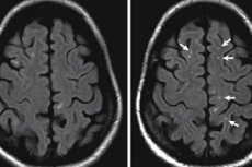
When diagnosing functional brain disorders after traumatic brain injury or stroke, examination of patients with signs of cerebral circulatory disorders, neurodegenerative diseases and various neurological disorders, neuropsychological methods are insufficient, and the key method is imaging - magnetic resonance imaging (MRI) of the brain, which reveals foci of gliosis.
The MRI picture of single supratentorial foci of gliosis consists of clearly limited areas of brain matter hyperintensity on T2-weighted images: small areas of diffuse enhancement are seen at the site of focal clusters of glial cells (on T1-weighted images these areas are hypointense, i.e. Light).
In this case, astrocytes are hypertrophied - with an increase in the size of cell nuclei and a decrease in the density of chromatin in them. [1]
Differential diagnosis
Differential diagnosis with subcortical or subependymal gliosis, glioma, leukoaraiosis, and periventricular leukomalacia is performed.
Who to contact?
Treatment of the supratentorial foci of gliosis.
Is it possible to treat gliosis foci in the supratentorial region? Gliosis is a process, and to date, therapeutic strategies have been sought to reduce the proliferation of neuroglia astrocytes and microglia.
Thus, tetracycline group antibiotic Minocycline inhibits microglia activation and suppresses astrocyte proliferation, but it has no effect on already formed foci. [2], [3]
Therefore, there is treatment of ischemic and hemorrhagic stroke, treatment of post-stroke condition or treatment of brain injury.
What methods are used to treat CNS disorders, read more in the publications:
Prevention
There are no specific medical recommendations regarding prophylactic measures to prevent pathologic proliferation or hypertrophy of brain neuroglia cells.
Forecast
The dependence of the outcome of pathology development on the localization of supratentorial foci of gliosis, their number, and the cause of death of neurons that have been replaced by neuroglia cells is obvious. In many cases, the prognosis is unfavorable with a high probability of patient disability.

