Medical expert of the article

New publications
The brain
Last reviewed: 04.07.2025

All iLive content is medically reviewed or fact checked to ensure as much factual accuracy as possible.
We have strict sourcing guidelines and only link to reputable media sites, academic research institutions and, whenever possible, medically peer reviewed studies. Note that the numbers in parentheses ([1], [2], etc.) are clickable links to these studies.
If you feel that any of our content is inaccurate, out-of-date, or otherwise questionable, please select it and press Ctrl + Enter.
The brain (encephalon) with the membranes surrounding it is located in the cavity of the brain section of the skull. In this regard, its convex upper-lateral surface in shape corresponds to the internal concave surface of the cranial vault. The lower surface - the base of the brain - has a complex relief corresponding to the shape of the cranial fossae of the internal base of the skull.
The weight of the adult human brain fluctuates between 1100 and 2000 g. The average length of the brain is 160-180 mm, the largest transverse dimension is 140 mm. The female brain is slightly shorter than the male brain on average. The average weight of the adult male brain is 1400 g, and that of a woman is 1200 g. The greatest brain weight is found in people aged 20 to 25 years. The average weight of the brachycephalic brain is heavier than that of the dolichocephalic brain.
There is no direct relationship between the weight of the brain and the intellectual capacity of a person. For example, the weight of the brain of the writer A. N. Turgenev is 2012 g, the poet Byron - 1807 g, the philosopher I. Kant - 1600 g, the poet I. F. Schiller - 1580 g, the doctor Broca - 1484 g, the doctor G. Dupuytren - 1437 g, the poet A. Dante - 1420 g, the artist A. Tiedemann - 1254 g. It is known that other people of outstanding intelligence had a brain with a comparatively small weight. The brain of idiots has an especially small weight, sometimes it does not even reach 300 g. Experience shows that spiritually more developed people often have a brain of a more significant weight. However, a high brain weight in no way indicates a higher spiritual development. At the same time, the weight of the brain must exceed a certain minimum norm so that mental functions can be performed correctly. For men, the minimum norm for the brain is considered to be 1000 g, and for women - 900 g. The spinal cord makes up about 2% of the brain weight and is equal to 34-38 g.
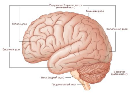
When examining a brain preparation, its three largest components are clearly visible: the cerebral hemispheres, the cerebellum, and the brain stem.
The cerebral hemispheres (hemispheriae cerebrales) in an adult are the most highly developed, largest, and functionally most important part of the central nervous system. The sections of the cerebral hemispheres cover all other parts of the brain.
The right and left hemispheres are separated from each other by a deep longitudinal fissure of the cerebrum (fissura longitudinalis cerebralis), which in the depths between the hemispheres reaches the large commissure of the brain, or corpus callosum. In the posterior sections, the longitudinal fissure connects with the transverse fissure of the cerebrum (fissura transversa cerebralis), which separates the cerebral hemispheres from the cerebellum.
On the upper lateral, medial and lower (basal) surfaces of the cerebral hemispheres there are deep and shallow grooves. Deep grooves divide each of the hemispheres into lobes of the cerebrum (lobi cerebrales). Shallow grooves are separated from each other by convolutions of the cerebrum (gyri cerebrales).
The inferior surface (facies inferior), or base of the brain, is formed by the ventral surfaces of the cerebral hemispheres, the cerebellum, and the ventral parts of the brain stem, which are most accessible here for viewing.
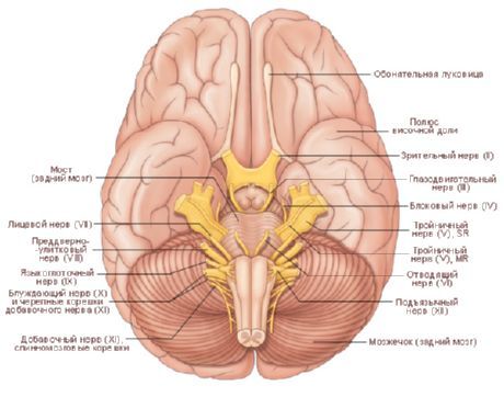
At the base of the brain, in the anterior sections formed by the lower surface of the frontal lobes of the cerebral hemispheres, one can find the olfactory bulbs (bulbi olfactorii). They look like small thickenings located on the sides of the longitudinal fissure of the cerebrum. 15-20 thin olfactory nerves (nn. olfactorii - I pair of cranial nerves) approach the ventral surface of each of the olfactory bulbs from the nasal cavity through openings in the ethmoid bone plate. When the brain is removed from the skull, the olfactory nerves are torn off and therefore are not visible on an isolated preparation.
From the olfactory bulb a cord extends backwards - the olfactory tract (tractus olfactorius). The posterior sections of the olfactory tract thicken and widen, forming the olfactory triangle (trigonum olfactorium). The posterior side of the olfactory triangle passes into a small area with a large number of small openings that remain after removal of the choroid. This is the anterior perforated substance (substantia perforata rostralis, s. anterior). Here, through the openings of the perforated substance, arteries penetrate deep into the brain. Medial to the perforated substance, closing the posterior sections of the longitudinal fissure of the cerebrum on the lower surface of the brain, is a thin, gray, easily torn terminal, or terminal, plate (lamina terminalis). The optic chiasma (chiasma opticum) is adjacent to this plate behind. It is formed by fibers that follow in the composition of the optic nerves (nn. opticum - II pair of cranial nerves), penetrating into the cranial cavity from the eye sockets. Two optic tracts (tractus opticus) depart from the optic chiasm in the posterolateral direction.
The grey tubercle (tuber cinereum) is adjacent to the posterior surface of the optic chiasm. The lower sections of the grey tubercle are elongated in the form of a tube tapering downwards, which is called the funnel (infundibulum). At the lower end of the funnel is a rounded formation - the pituitary gland (hypophysis), an endocrine gland. The pituitary gland is located in the cranial cavity in the fossa sella turcica and when the brain preparation is removed from the skull, it remains in this depression, breaking away from the funnel.
Two white spherical elevations, the mammillary bodies (corpora mamillaria), adjoin the gray tubercle at the back. Behind the optic tracts, two longitudinal white ridges are visible - the cerebral peduncles (pedunculi cerebri), between which there is a depression - the interpeduncular fossa (fossa interpeduncularis), limited in front by the mammillary bodies. The bottom of this fossa is formed by the posterior perforated substance (substantia perforata interpeduncularis posterior), through the openings of which the arteries that feed the brain penetrate into it. On the medial surfaces of the cerebral peduncles facing each other, the roots of the right and left oculomotor nerves (nn. oculomotorius - III pair of cranial nerves) are visible. The lateral surfaces of the cerebral peduncles are encircled by the trochlear nerves (nn. trochleares - IV pair of cranial nerves), the roots of which exit the brain not at its base, as in all the other 11 pairs of cranial nerves, but on the dorsal surface, behind the lower colliculi of the roof of the midbrain, on the sides of the frenulum of the superior medullary velum.
The cerebral peduncles emerge from the upper sections of the broad transverse ridge, which is designated as the bridge (pons). The lateral sections of the bridge continue into the cerebellum, forming the paired middle cerebellar peduncle (pedunculus cerebellaris medius).
At the border between the pons and the middle cerebellar peduncles, the root of the trigeminal nerve (n. trigeminus - V pair of cranial nerves) can be seen on each side.
Below the bridge are the anterior sections of the medulla oblongata, which are represented by medially located pyramids separated from each other by the anterior median fissure. Lateral to the pyramid is a rounded elevation - the olive. At the border of the bridge and the medulla oblongata, on the sides of the anterior median fissure, the roots of the abducens nerve (n. abducens - VI cranial nerve) emerge from the brain. Even more laterally, between the middle cerebellar peduncle and the olive, on each side are successively located the roots of the facial nerve (n. facialis - VII cranial nerve), and the vestibulocochlearis nerve (n. vestibulocochlearis - VIII cranial nerve). Dorsal to the olive, in an inconspicuous groove, the roots of the following cranial nerves pass from front to back: glossopharyngeal (n. glossopharyngeus - IX nerve), vagus (n. vagus - X nerve) and accessory (n. accessorius - XI nerve). The roots of the accessory nerve also extend from the spinal cord in its upper part - these are the spinal roots (radices spinales; spinal part, pars spinalis). In the groove separating the pyramid from the olive, there are roots of the hypoglossal nerve (n. hypogosus - XII pair of cranial nerves).
On a median sagittal section of the brain, drawn along the longitudinal fissure of the cerebrum, the medial surface of the cerebral hemisphere, some structures of the brainstem (truncus encephalicus) and the cerebellum are visible.
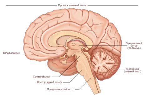
The vast medial surface of the cerebral hemispheres overhangs the much smaller cerebellum and brainstem. On this surface, as on other surfaces, there are furrows that separate the convolutions of the cerebrum from each other.
The frontal, parietal and occipital lobes of each hemisphere are separated from the large cerebral commissure, the corpus callosum, which is clearly visible in the median section, by the corpus callosum groove (sulcus corporis callosi). The middle part of the corpus callosum is called the trunk (truncus). Its anterior sections, bending downwards, form the knee (genu). Even further downwards, the knee of the corpus callosum becomes thinner and is called the beak (rostrum), which continues downwards into the terminal plate (lamina terminalis). The latter, as noted, fuses with the anterior surface of the optic chiasm. The posterior sections of the corpus callosum are noticeably thickened and end freely in the form of a ridge (splenium).
Under the corpus callosum there is a thin white plate - the fornix. Gradually moving away from the corpus callosum and forming an arcuate bend forward and downwards, the body of the fornix continues into a column (columna) of the fornix. The lower part of each column of the fornix first approaches the terminal plate, and then moves laterally and is directed back, ending in the mammillary body. Between the columns of the fornix at the back and the terminal plate at the front there is a transverse bundle of nerve fibers that have the appearance of a white oval in section - this is the anterior (white) commissure (commissure rostralis, s. anterior). The commissure, like the transverse fibers of the corpus callosum, connects the hemispheres of the brain, its anterior sections, with each other.
The area bounded above and in front by the corpus callosum, below by its beak, terminal plate and anterior commissure, and behind by the column of the fornix, is occupied by a thin sagittally oriented plate of the medulla - the transparent septum (septum pellucidum).
All of the above formations belong to the telencephalon. The structures located below, with the exception of the cerebellum, belong to the brainstem. The most anterior sections of the brainstem are formed by the right and left thalamuses - this is the posterior thalamus (thalamus dorsalis). The thalamus is located below the body of the fornix and the corpus callosum and behind the column of the fornix. In the median section, only the medial surface of the posterior thalamus is distinguishable. The interthalamic fusion (adhesio interthalamica) is distinguished on it. The medial surface of each posterior thalamus laterally limits the slit-like, vertically located cavity of the third ventricle. Between the anterior end of the thalamus and the column of the fornix is the interventricular foramen (foramen interventriculare), through which the lateral ventricle of the cerebral hemisphere communicates with the cavity of the third ventricle. In the posterior direction from the interventricular opening, the hypothalamic (subthalamic) groove (sulcus hypothalamicus) extends, bending around the thalamus from below. The formations located below this groove belong to the hypothalamus. These are the optic chiasm, the gray tubercle, the infundibulum, the pituitary gland and the mammillary bodies - structures that participate in the formation of the bottom of the third ventricle.
Above and behind the thalamus, under the splenium of the corpus callosum, is the pineal body (corpus pineale), which is an endocrine gland. The anterior-inferior parts of the pineal body are fused with a thin transverse cord, rounded on the sagittal section. This cord is the epithalamic commissure (commissura epithalamica). The thalamus (thalamus), hypothalamus, third ventricle, and pineal body belong to the diencephalon.
Caudal to the thalamus are the structures related to the midbrain (mesencephalon). Below the pineal body is the roof of the midbrain (tectum mesencephalicum), consisting of two superior and two inferior colliculi. Ventral to the roof of the midbrain is the peduncle of the brain (pedunculus cerebri), separated from the roof by the midbrain aqueduct.
The aqueduct of the midbrain (aqueductus mesencephali) connects the cavities of the third and fourth ventricles. Even more posteriorly are the median sections of the pons and cerebellum, which belong to the hindbrain (metencephalon), and the section of the medulla oblongata (medulla oblongata). The cavity of these parts of the brain is the fourth ventricle (ventriculus quartos). The floor of the fourth ventricle is formed by the dorsal surface of the pons and medulla oblongata, which forms the rhomboid fossa (fossa rhomboidea) on the whole brain. A thin plate of white matter, which stretches from the cerebellum to the roof of the midbrain, is called the superior medullary velum (velum medullare rostralis, s. superius). From the lower surface of the cerebellum back to the medulla oblongata, extends the inferior medullary velum (velum medullare caudale, s. inferius).
There are 5 parts of the brain that develop from five cerebral vesicles:
- end brain;
- diencephalon;
- midbrain;
- hindbrain;
- the medulla oblongata, which at the level of the foramen magnum passes into the spinal cord.
 [ 1 ]
[ 1 ]
Functions of the brain
The human brain performs many important functions and is the central part of the nervous system. Here are the main functions of the brain:
Cognitive functions:
- Thinking: The brain processes information, allowing a person to solve problems, make decisions, and reason.
- Memory: The brain is involved in the formation and storage of long-term and short-term memory.
- Attention and concentration: It helps you focus on specific tasks and filter information.
- Language and Communication: The brain controls language skills and the ability to communicate.
Sensory and motor functions:
- Senses: The brain processes information from the senses such as sight, hearing, smell, taste and touch.
- Movement: It controls motor skills and coordination of movements.
Regulation of internal organs:
- The brain controls important functions such as breathing, heartbeat, temperature regulation and digestion.
Emotions and behavior:
- It is involved in the formation and regulation of emotions, mood and behavior.
Consciousness and perception of the surrounding world:
- The brain is responsible for awareness of the surrounding world and the formation of consciousness.
Preservation of vital functions:
- It controls autonomic functions such as regulating blood pressure, blood glucose levels, and others.
Training and adaptation:
- The brain facilitates learning and adaptation to new information and situations.
Response to stress and danger:
- It responds to stressful situations and danger by activating the fight or flight response.
The brain is a complex and multifaceted structure consisting of various areas and subsystems, each of which is responsible for certain functions. Its work depends on the correct functioning of millions of neurons and their interaction with each other.
Development of the brain in the fetus
Fetal brain development occurs gradually and goes through several key stages throughout pregnancy. Here's a quick rundown:
- 1-2 weeks: At the earliest stage of pregnancy, the egg is fertilized and the zygote is formed. At this time, the process of formation of the neuronal plate, the initial structure of the future nervous tissue, begins.
- 3-4 weeks: The neural plate begins to close and form the neural tube. Closure of the anterior and posterior neuropores also occurs during this period, which is critical to preventing neural tube defects.
- 5-8 weeks: The neural tube differentiates into the different parts of the brain, including the cerebellum, diencephalon, hindbrain, and brainstem. Neurons begin to migrate to their future locations in the brain.
- 9-12 weeks: At this stage, intensive proliferation and migration of neurons occurs. The brain begins to acquire a more complex structure, and connections between neurons begin to form.
- 13-16 weeks: The brain becomes more complex and the cerebral cortex, which plays a key role in cognitive functions, begins to actively develop.
- 17-20 weeks: At this time, the folds and grooves on the surface of the brain begin to form. The brain begins to control some functions, such as fetal movements.
- 21-24 weeks: The cerebral cortex is developing rapidly and many neural connections begin to form.
- 25-28 weeks: The brain continues to grow and develop, and the fetus begins to respond to external stimuli.
- 29-32 weeks: Neural connections become more complex, and the brain begins to prepare to control the functions it will perform after birth.
- 33-40 weeks: During the last weeks of pregnancy, the brain continues to develop and strengthen its functions in preparation for birth and life outside the womb.
This is a general overview of fetal brain development week by week. It is important to remember that every pregnancy and fetus is unique, and development may vary slightly from case to case. Fetal brain development is a complex and fascinating process that demonstrates the body's amazing ability to self-regulate and self-heal.
Brain diseases
The brain can be affected by a variety of diseases and conditions. Here are some of the most common diseases and conditions that can affect the brain:
- Hydrocephalus: A condition in which the ventricles of the brain become filled with excess cerebrospinal fluid.
- Migraine: Paroxysmal headaches that are often accompanied by aura, photophobia and nausea.
- Epilepsy: A neurological disorder characterized by seizures.
- Stroke: An acute interruption of blood supply to the brain that can lead to impaired brain function.
- Head injuries: Includes bruises, concussions, and other brain injuries.
- Brain tumors: Malignant and benign tumors that develop inside the skull.
- Alzheimer's disease: A progressive neurodegenerative disease associated with cognitive decline.
- Parkinsonism: A group of neurological disorders characterized by impaired motor skills and jerky movements.
- Multiple sclerosis: An autoimmune disease that attacks the myelin of nerves and causes a variety of neurological symptoms.
- CP (cerebral palsy): A group of neurological disorders that occur in early childhood due to damage to the brain.
- Dementia: A general term for conditions characterized by a deterioration in a patient's cognitive functions and abilities.
- Cerebral hypoxia and ischemia: Lack of oxygen or lack of blood supply to the brain, which can cause damage to brain cells.
- Inflammatory diseases of the brain: For example, meningitis and encephalitis, which are characterized by inflammation of the meninges and brain tissue.
- Neurodegenerative diseases: For example, Huntington's disease, Parkinson's disease, etc.
- Congenital and developmental brain abnormalities: Abnormalities a child is born with can affect the development and functioning of the brain.
This is just a small list of brain diseases. Each of these conditions has its own unique symptoms, causes, and treatments, and diagnosis and treatment should be done under the guidance of qualified medical professionals.
Where does it hurt?
What's bothering you?
What do need to examine?