Medical expert of the article

New publications
The medulla oblongata
Last reviewed: 04.07.2025

All iLive content is medically reviewed or fact checked to ensure as much factual accuracy as possible.
We have strict sourcing guidelines and only link to reputable media sites, academic research institutions and, whenever possible, medically peer reviewed studies. Note that the numbers in parentheses ([1], [2], etc.) are clickable links to these studies.
If you feel that any of our content is inaccurate, out-of-date, or otherwise questionable, please select it and press Ctrl + Enter.
The medulla oblongata (s. myelencephalon) is located between the hindbrain and the spinal cord. The upper border of the medulla oblongata on the ventral surface of the brain runs along the lower edge of the pons. On the dorsal surface, this border corresponds to the medullary stripes of the fourth ventricle, which divide the bottom of the fourth ventricle into upper and lower parts. The border between the medulla oblongata and the spinal cord corresponds to the level of the foramen magnum or the place where the upper part of the roots of the first pair of spinal nerves exits the brain.
The upper sections of the medulla oblongata are somewhat thicker than the lower ones. In this connection, the medulla oblongata takes the form of a truncated cone or bulb, for the resemblance to which it is also called a bulb (bulbus). The length of the medulla oblongata of an adult is on average 25 mm.
The medulla oblongata has a ventral, dorsal, and two lateral surfaces, which are separated by grooves. The grooves of the medulla oblongata are a continuation of the grooves of the spinal cord and have the same names. These are the anterior median fissure (fissura mediana ventralis, s. anterior); posterior median groove (sulcus medianus dorsalis, s. posterior); anterolateral groove (sulcus ventrolateralis, s. anterolateralis); posterolateral groove (sulcus dorsolateralis, s. posterolateral). On both sides of the anterior median fissure on the ventral surface of the medulla oblongata there are convex ridges, gradually narrowing downwards - pyramids (pyramides). In the lower part of the medulla oblongata, the bundles of fibers that make up the pyramids cross to the opposite side and enter the lateral funiculi of the spinal cord. This transition of fibers is called the decussation of the pyramids (decussatio pyramidum, s. decussatio motoria; motor decussation). The decussation site also serves as an anatomical border between the medulla oblongata and the spinal cord. To the side of each pyramid of the medulla oblongata is an oval elevation - the olive (oliva), which is separated from the pyramid by the anterolateral groove. In this groove, the roots of the hypoglossal nerve (XII pair) emerge from the medulla oblongata.
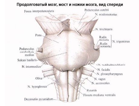
On the dorsal surface, on the sides of the posterior median groove, the thin and cuneate fascicles of the posterior funiculi of the spinal cord end in thickenings, separated from each other by the posterior intermediate groove. The thin fascicle (fasciculus gracilis), lying more medially, expands to form the tubercle of the thin nucleus (tuberculum gracile). The cuneate fascicle (fasciculus cuneatus) is located more laterally, which forms the tubercle of the cuneate nucleus (tuberculum cuneatum) on the side of the tubercle of the thin fascicle. Dorsal to the olive, the roots of the glossopharyngeal, vagus and accessory nerves (IX, X and XI pairs) emerge from the posterolateral groove of the medulla oblongata - the retroolive groove (sulcus retroolivaris).
The dorsal part of the lateral funiculus widens slightly upward. Here, fibers from the cuneate and thin nuclei join it. Together, they form the inferior cerebellar peduncle, also called the cord-shaped body. The surface of the medulla oblongata, limited below and laterally by the inferior cerebellar peduncles, participates in the formation of the rhomboid fossa, which is the bottom of the fourth ventricle.
A cross-section through the medulla oblongata at the level of the olives shows clusters of white and gray matter. In the lower lateral sections are the right and left inferior olivary nuclei (nuclei olivares caudales [inferiores]). They are curved in such a way that their gates face medially and upward. Somewhat above the inferior olivary nuclei is the reticular formation (formatio reticularis), formed by the interweaving of nerve fibers and the nerve cells lying between them and their clusters in the form of small nuclei. Between the inferior olivary nuclei is the so-called interolive layer, represented by the internal arcuate fibers (fibrae arcuatae internae) - processes of cells lying in the thin and cuneate nuclei. These fibers form the medial loop (lemniscus medialis). The fibers of the medial lemniscus belong to the proprioceptive pathway of the cortical direction and form the decussatio lemniscorum medianum in the medulla oblongata. In the upper lateral parts of the medulla oblongata, the right and left inferior cerebellar peduncles are visible in section. The fibers of the anterior spinocerebellar and rubrospinal tracts pass somewhat ventrally. In the central part of the medulla oblongata, on the sides of the anterior median fissure, are the pyramids. The medial longitudinal fasciculus (fasciculus longitudinalis medialis [posterior]) is located above the decussatio lemniscorum medianum.
The medulla oblongata contains the nuclei of the IX, X, XI and XII pairs of cranial nerves, which participate in the innervation of the internal organs and derivatives of the gill apparatus. The ascending pathways to other parts of the brain also pass here. The ventral parts of the medulla oblongata are represented by descending motor pyramidal fibers. Dorsolaterally, ascending pathways pass through the medulla oblongata, connecting the spinal cord with the cerebral hemispheres, the brain stem and the cerebellum. In the medulla oblongata, as in some other parts of the brain, there is a reticular formation, as well as such vital centers as the centers of blood circulation, respiration and digestion.
 [
[ What do need to examine?
How to examine?