Medical expert of the article

New publications
Basal (subcortical) nuclei
Last reviewed: 07.07.2025

All iLive content is medically reviewed or fact checked to ensure as much factual accuracy as possible.
We have strict sourcing guidelines and only link to reputable media sites, academic research institutions and, whenever possible, medically peer reviewed studies. Note that the numbers in parentheses ([1], [2], etc.) are clickable links to these studies.
If you feel that any of our content is inaccurate, out-of-date, or otherwise questionable, please select it and press Ctrl + Enter.
Basal (subcortical) nuclei and white matter of the telencephalon
In addition to the cortex, which forms the surface layers of the end brain, the gray matter in each of the cerebral hemispheres is located in the form of separate nuclei, or nodes. These nodes are located in the thickness of the white matter, closer to the base of the brain. The accumulations of gray matter, due to their position, are called basal (subcortical, central) nuclei (nuclei basales), or nodes. The basal nuclei of the hemispheres include the striatum, consisting of the caudate and lenticular nuclei, the enclosure, and the amygdala.
The corpus striatum gets its name from the fact that on horizontal and frontal sections of the brain it looks like alternating stripes of gray and white matter. The caudate nucleus (nucleus caudatus) is located most medially and in front. It is located in front of the thalamus, from which (on a horizontal section) it is separated by a strip of white matter - the anterior leg of the internal capsule. The anterior part of the caudate nucleus is thickened and forms the head (caput), which makes up the lateral wall of the anterior horn of the lateral ventricle. Situated in the frontal lobe of the hemisphere, the head of the caudate nucleus adjoins the anterior perforated substance. At this point, the head of the caudate nucleus connects with the lentiform nucleus. Tapering posteriorly, the head continues into a thinner body (corpus), which lies in the area of the bottom of the central part of the lateral ventricle and is separated from the thalamus by a terminal strip of white matter. The posterior part of the caudate nucleus - the tail (cauda) gradually becomes thinner, bends downwards, and participates in the formation of the upper wall of the inferior horn of the lateral ventricle. The tail reaches the amygdala, which lies in the anteromedial parts of the temporal lobe (behind the anterior perforated substance). Laterally to the head of the caudate nucleus is a layer of white matter - the anterior leg (thigh) of the internal capsule, separating this nucleus from the lenticular one.
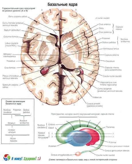
The lentiform nucleus (nucleus lentiformis), named for its resemblance to a lentil, is located lateral to the thalamus and caudate nucleus. The posterior leg (hip) of the internal capsule separates the lentiform nucleus from the thalamus. The lower surface of the anterior part of the lentiform nucleus is adjacent to the anterior perforated substance and connects with the caudate nucleus. The medial part of the lentiform nucleus on a horizontal section of the brain narrows and faces the genu of the internal capsule, located on the border of the thalamus and the head of the caudate nucleus.
The lateral surface of the lentiform nucleus is convex and faces the base of the insular lobe of the cerebral hemisphere. In the frontal section of the brain, the lentiform nucleus has the shape of a triangle, the apex of which faces the medial side, and the base - the lateral side. Two parallel vertical layers of white matter, located almost in the sagittal plane, divide the lentiform nucleus into three parts. The most laterally located is the putamen, which has a darker color. Medial to the putamen are two light brain plates - medial and lateral (laminae medullares medialis et lateralis), which are united by the common name "pale blinder" (globus pallidus).
The medial plate is called the medial globus pallidus medialis, the lateral plate is called the lateral globus pallidus lateralis. The caudate nucleus and putamen are phylogenetically newer formations (neosti latum, s. striatum). The globus pallidus is an older formation (paleostriatum, s. pallidum).
The claustrum is located in the white matter of the hemisphere, to the side of the putamen, between the latter and the cortex of the insular lobe. The claustrum has the appearance of a thin vertical plate of gray matter. It is separated from the putamen by a layer of white matter - the external capsule (capsula externa), and from the cortex of the insula - by the same layer, called the "most external capsule" (capsula extria).
The amygdala (corpus amygdaloideum) is located in the white matter of the temporal lobe of the hemisphere, approximately 1.5-2.0 cm posterior to the temporal pole.
 [
[ What do need to examine?
How to examine?