Medical expert of the article
New publications
The blood-brain barrier
Last reviewed: 07.07.2025

All iLive content is medically reviewed or fact checked to ensure as much factual accuracy as possible.
We have strict sourcing guidelines and only link to reputable media sites, academic research institutions and, whenever possible, medically peer reviewed studies. Note that the numbers in parentheses ([1], [2], etc.) are clickable links to these studies.
If you feel that any of our content is inaccurate, out-of-date, or otherwise questionable, please select it and press Ctrl + Enter.
The blood-brain barrier is extremely important for ensuring homeostasis of the brain, but many questions concerning its formation have not yet been fully clarified. But it is already clear that the BBB is the most differentiated, complex, and dense histohematic barrier. Its main structural and functional unit is the endothelial cells of the brain capillaries.
The metabolism of the brain, like no other organ, depends on substances entering with the bloodstream. Numerous blood vessels that ensure the functioning of the nervous system are distinguished by the fact that the process of penetration of substances through their walls is selective. Endothelial cells of the capillaries of the brain are connected to each other by continuous tight contacts, so substances can pass only through the cells themselves, but not between them. Glia cells, the second component of the blood-brain barrier, are adjacent to the outer surface of the capillaries. In the vascular plexuses of the ventricles of the brain, the anatomical basis of the barrier are epithelial cells, also tightly connected to each other. At present, the blood-brain barrier is considered not as an anatomical and morphological, but as a functional formation capable of selectively passing, and in some cases delivering various molecules to nerve cells using active transport mechanisms. Thus, the barrier performs regulatory and protective functions
There are structures in the brain where the blood-brain barrier is weakened. These are primarily the hypothalamus, as well as a number of structures at the bottom of the 3rd and 4th ventricles - the most posterior field (area postrema), the subfornical and subcommissural organs, and the pineal body. The integrity of the BBB is disrupted in ischemic and inflammatory brain lesions.
The blood-brain barrier is considered to be fully formed when the properties of these cells satisfy two conditions. First, the rate of liquid-phase endocytosis (pinocytosis) in them must be extremely low. Second, specific tight junctions must form between the cells, which are characterized by very high electrical resistance. It reaches values of 1000-3000 Ohm/cm2 for capillaries of the pia mater and from 2000 to 8000 0 m/cm2 for intraparenchymal cerebral capillaries. For comparison: the average value of transendothelial electrical resistance of skeletal muscle capillaries is only 20 Ohm/cm2.
The permeability of the blood-brain barrier for most substances is largely determined by their properties, as well as the ability of neurons to synthesize these substances independently. Substances that can overcome this barrier include, first of all, oxygen and carbon dioxide, as well as various metal ions, glucose, essential amino acids and fatty acids necessary for normal brain function. Glucose and vitamins are transported using carriers. At the same time, D- and L-glucose have different rates of penetration through the barrier - the former is more than 100 times higher. Glucose plays a major role both in the energy metabolism of the brain and in the synthesis of a number of amino acids and proteins.
The leading factor determining the functioning of the blood-brain barrier is the level of metabolism of nerve cells.
Provision of neurons with necessary substances is carried out not only by means of blood capillaries approaching them, but also thanks to the processes of the soft and arachnoid membranes, through which cerebrospinal fluid circulates. Cerebrospinal fluid is located in the cranial cavity, in the ventricles of the brain and in the spaces between the membranes of the brain. In humans, its volume is about 100-150 ml. Thanks to the cerebrospinal fluid, the osmotic balance of nerve cells is maintained and metabolic products toxic to nerve tissue are removed.

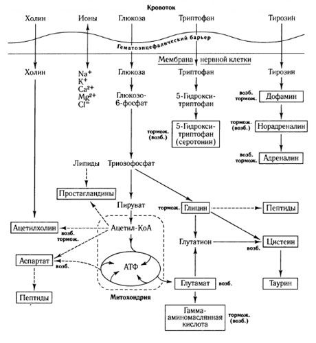
Pathways of mediator exchange and the role of the blood-brain barrier in metabolism (according to: Shepherd, 1987)
The passage of substances through the blood-brain barrier depends not only on the permeability of the vascular wall to them (molecular weight, charge and lipophilicity of the substance), but also on the presence or absence of an active transport system.
The stereospecific insulin-independent glucose transporter (GLUT-1), which ensures the transfer of this substance across the blood-brain barrier, is abundant in endothelial cells of brain capillaries. The activity of this transporter can ensure the delivery of glucose in an amount 2-3 times greater than that required by the brain under normal conditions.
Characteristics of the transport systems of the blood-brain barrier (according to: Pardridge, Oldendorf, 1977)
Transportable |
Preferential substrate |
Km, mm |
Vmax |
Hexoses |
Glucose |
9 |
1600 |
Monocarboxylic |
Lactate |
1.9 |
120 |
Neutral |
Phenylalanine |
0.12 |
30 |
Essential |
Lysine |
0.10 |
6 |
Amines |
Choline |
0.22 |
6 |
Purines |
Adenine |
0.027 |
1 |
Nucleosides |
Adenosine |
0,018 |
0.7 |
Children with impaired functioning of this transporter experience a significant decrease in the level of glucose in the cerebrospinal fluid and disturbances in the development and functioning of the brain.
Monocarboxylic acids (L-lactate, acetate, pyruvate) and ketone bodies are transported by separate stereospecific systems. Although their transport intensity is lower than that of glucose, they are an important metabolic substrate in neonates and during starvation.
Choline transport into the central nervous system is also mediated by the transporter and can be regulated by the rate of acetylcholine synthesis in the nervous system.
Vitamins are not synthesized by the brain and are supplied from the blood using special transport systems. Despite the fact that these systems have a relatively low transport activity, under normal conditions they can ensure the transport of the amount of vitamins necessary for the brain, but their deficiency in food can lead to neurological disorders. Some plasma proteins can also penetrate the blood-brain barrier. One of the ways of their penetration is receptor-mediated transcytosis. This is how insulin, transferrin, vasopressin and insulin-like growth factor penetrate the barrier. Endothelial cells of brain capillaries have specific receptors for these proteins and are able to endocytose the protein-receptor complex. It is important that as a result of subsequent events, the complex disintegrates, the intact protein can be released on the opposite side of the cell, and the receptor can again be integrated into the membrane. For polycationic proteins and lectins, transcytosis is also a way of penetrating the BBB, but it is not associated with the work of specific receptors.
Many neurotransmitters present in the blood are not able to penetrate the BBB. Thus, dopamine does not have this ability, while L-DOPA penetrates the BBB using the neutral amino acid transport system. In addition, capillary cells contain enzymes that metabolize neurotransmitters (cholinesterase, GABA transaminase, aminopeptidases, etc.), drugs and toxic substances, which ensures the protection of the brain not only from neurotransmitters circulating in the blood, but also from toxins.
The work of the BBB also involves carrier proteins that transport substances from the endothelial cells of the brain capillaries into the blood, preventing their penetration into the brain, for example, b-glycoprotein.
During ontogenesis, the rate of transport of various substances through the BBB changes significantly. Thus, the rate of transport of b-hydroxybutyrate, tryptophan, adenine, choline, and glucose in newborns is significantly higher than in adults. This reflects the relatively higher need of the developing brain for energy and macromolecular substrates.


 [
[