Medical expert of the article

New publications
Lung cancer
Last reviewed: 12.07.2025

All iLive content is medically reviewed or fact checked to ensure as much factual accuracy as possible.
We have strict sourcing guidelines and only link to reputable media sites, academic research institutions and, whenever possible, medically peer reviewed studies. Note that the numbers in parentheses ([1], [2], etc.) are clickable links to these studies.
If you feel that any of our content is inaccurate, out-of-date, or otherwise questionable, please select it and press Ctrl + Enter.
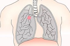
Lung cancer is a malignant tumor of the lung, usually classified as small cell or non-small cell. Cigarette smoking is the main risk factor for most types of the tumor. Symptoms include cough, chest discomfort, and, less commonly, hemoptysis, but many patients are asymptomatic and some develop metastatic lesions. Diagnosis is suspected by chest radiography or CT scan and confirmed by biopsy. Treatment includes surgery, chemotherapy, and radiation therapy. Despite advances in therapy, the prognosis is poor and attention should be focused on early detection and prevention.
Causes lung cancer
Cigarette smoking, including passive smoking, is the most important cause of lung cancer. The risk depends on age, intensity, and duration of smoking; the risk declines after smoking cessation but probably never returns to baseline. In nonsmokers, the most important environmental risk factor is exposure to radon, a breakdown product of naturally occurring radium and uranium. Occupational hazards include exposure to radon (uranium miners); asbestos (construction and demolition workers, plumbers, shipbuilders, and auto mechanics); quartz (miners and sandblasters); arsenic (copper smelters, pesticide manufacturers, and plant protection product manufacturers); chromium derivatives (stainless steel manufacturers and pigment manufacturers); nickel (battery manufacturers and stainless steel manufacturers); chloromethyl ethers; beryllium and coke oven emissions (in steel workers), account for a small number of cases each year. The risk of malignant neoplasms of the respiratory system is higher when occupational hazards and cigarette smoking are combined than when either is present alone. COPD and pulmonary fibrosis may increase the risk; beta-carotene supplements may increase the risk in smokers. Air pollution and cigar smoke contain carcinogens, but their role in the development of lung cancer has not been proven.
Symptoms lung cancer
Approximately 25% of all cases of the disease are asymptomatic and are discovered incidentally during chest examination. Symptoms of lung cancer consist of local manifestations of the tumor, regional spread, and metastases. Paraneoplastic syndromes and general manifestations can occur at any stage.
Local symptoms include cough and, less commonly, dyspnea due to airway obstruction, post-obstructive atelectasis, and lymphatic spread. Fever may occur with the development of post-obstructive pneumonia. Up to half of patients complain of vague or localized chest pain. Hemoptysis is less common and blood loss is minimal, except in rare cases when the tumor ruptures a major artery, causing massive hemorrhage and death due to asphyxia.
Regional spread may cause pleuritic pain or dyspnea due to the development of pleural effusion, dysphonia due to tumor invasion of the recurrent laryngeal nerve, and dyspnea and hypoxia due to diaphragmatic paralysis when the phrenic nerve is involved.
Compression of or invasion of the superior vena cava (superior vena cava syndrome) may cause headache or a feeling of fullness in the head, swelling of the face or upper extremities, shortness of breath, and flushing (plethora) when supine. Manifestations of superior vena cava syndrome include swelling of the face and upper extremities, distention of the jugular and subcutaneous veins of the face and upper torso, and flushing of the face and torso. Superior vena cava syndrome is more common in patients with the small cell type.
Apical neoplasms, usually non-small cell, may invade the brachial plexus, pleura, or ribs, causing shoulder and upper extremity pain and weakness or atrophy of one arm (Pancoast tumor). Horner's syndrome (ptosis, miosis, anophthalmos, and anhidrosis) occurs when the paravertebral sympathetic chain or cervical stellate ganglion is involved. Pericardial extension may be asymptomatic or result in constrictive pericarditis or cardiac tamponade. Rarely, esophageal compression results in dysphagia.
Metastases always eventually cause manifestations related to their location. Liver metastases cause gastrointestinal symptoms and, ultimately, liver failure. Brain metastases result in behavioral disturbances, amnesia, aphasia, seizures, paresis or paralysis, nausea and vomiting, and, ultimately, coma and death. Bone metastases cause intense pain and pathological fractures. Malignant neoplasms of the respiratory system often metastasize to the adrenal glands, but rarely lead to adrenal insufficiency.
Paraneoplastic syndromes are not caused directly by the cancer. Common paraneoplastic syndromes in patients include hypercalcemia (caused by tumor production of parathyroid hormone-related protein), syndrome of inappropriate antidiuretic hormone secretion (SIADH), clubbing of the fingers with or without hypertrophic osteoarthropathy, hypercoagulability with migratory superficial thrombophlebitis (Trousseau syndrome), myasthenia gravis (Eaton-Lambert syndrome), and a variety of neurologic syndromes including neuropathies, encephalopathies, encephalitides, myelopathies, and cerebellar lesions. The mechanism for the development of neuromuscular syndromes involves tumor expression of autoantigens with the formation of autoantibodies, but the cause of most others is unknown.
General symptoms usually include weight loss, malaise and are sometimes the first signs of malignancy.
Where does it hurt?
What's bothering you?
Stages
| Primary tumor | |
| Tis | Carcinoma in situ |
| T1 | Tumor < 3 cm without invasion, located proximal to the lobar bronchus (i.e. not in the main bronchus) |
| T2 | Tumor with any of the following features: >3cm Involves the main bronchus >2 cm distal to the carina Invades the visceral pleura Atelectasis or post-obstructive pneumonia that extends apically but does not involve the entire lung |
| TZ | Tumor of any size with any of the following features: Invades the chest wall (including superior sulcus lesions), diaphragm, mediastinal pleura, or parietal pericardium Involves a main bronchus < 2 cm distal to the carina but without carinal involvement Atelectasis or post-obstructive pneumonia of the entire lung |
| T4 | Tumor of any size with any of the following features: Invades the mediastinum, heart, great vessels, trachea, esophagus, vertebral body, carina Malignant pleural or pericardial effusion Satellite nodules of neoplasm within the same lobe as the primary tumor |
| Regional lymph nodes (N) | |
| N0 | No metastases to regional lymph nodes |
| N1 | Unilateral metastases to peribronchial lymph nodes and/or lymph nodes of the lung root and intrapulmonary lymph nodes located on the direct path of spread of the primary neoplasm |
| N2 | Unilateral metastases to mediastinal and/or subcarinal lymph nodes |
| N3 | Metastases to contralateral mediastinal nodes, contralateral root nodes, scalene muscle of the corresponding side or contralateral or supraclavicular lymph nodes |
| Distant metastases (M) | |
| M0 | No distant metastases |
| M1 | Distant metastases are present (including metastatic nodes in the lobes of the corresponding side but other than the primary tumor) |
| Stage 0 Tis IA T1 N0 M0 IB T2 N0 M0 IIA T1 N1 M0 |
Stage IIB T2N1 M0 or T3 N0 M0 IIIA T3 N1 M0 or TI-3 N2 M0 IIIB Any T N M0 or T4 any N M0 IV any T any N M1 |
Forms
Malignant
- Carcinoma
- Small cell
- Oat cell
- Transitional cell
- Mixed
- Non-small cell
- Adenocarcinoma
- Acinar
- Bronchioloalveolar
- Papillary
- Solid
- Adenosquamous
- Large cell
- Clear cell
- Giant cell
- Squamous cell
- Spindle cell
- Bronchial gland carcinoma
- Adenoid cystic
- Mucoepidermoid
- Carcinoid
- Lymphoma
- Primary pulmonary Hodgkin's disease
- Primary pulmonary non-Hodgkin's disease
Benign
- Laryngotracheobronchial
- Adenoma
- Hamartoma
- Myoblastoma
- Papilloma
- Parenchymal
- Fibroma
- Hamartoma
- Leiomyoma
- Lipoma
- Neurofibroma/schwannoma
- Sclerosing hemangioma
Malignant transformation of respiratory epithelial cells requires long-term contact with carcinogenic substances and accumulation of multiple genetic mutations. Mutations in genes that stimulate cell growth (K-RAS, MYC), encode growth factor receptors (EGFR, HER2/neu) and inhibit apoptosis (BCL-2) contribute to the proliferation of pathological cells. Mutations that inhibit tumor suppressor genes (p53, APC) have the same effect. When these mutations accumulate sufficiently, malignant neoplasms of the respiratory organs develop.
Lung cancer is usually divided into small cell lung cancer (SCLC) and non-small cell lung cancer (NSCLC). Small cell lung cancer is a very aggressive tumor, almost always occurs in smokers, and causes widespread metastasis in 60% of patients by the time of diagnosis. Symptoms of the non-small cell type are more variable and depend on the histological type.
Complications and consequences
Treatment of malignant pleural effusions begins with thoracentesis. Asymptomatic effusions do not require therapy; symptomatic effusions that recur despite multiple thoracenteses are drained through a chest tube. Injection of talc (or sometimes tetracycline or bleomycin) into the pleural space (a procedure called pleurodesis) causes pleural sclerosis, eliminates the pleural cavity, and is effective in more than 90% of cases.
Treatment of superior vena cava syndrome is similar to that of lung cancer: chemotherapy, radiation therapy, or both. Glucocorticoids are commonly used, but their effectiveness is unproven. Apical tumors are treated with surgery with or without preoperative radiation therapy or radiation therapy with or without adjuvant chemotherapy. Treatment of paraneoplastic syndromes depends on the specific situation.
Diagnostics lung cancer
The first investigation is a chest radiograph. It can clearly show specific abnormalities, such as single or multiple infiltrates or an isolated pulmonary nodule, or more subtle changes, such as thickened interlobar pleura, widened mediastinum, tracheobronchial narrowing, atelectasis, non-resolving parenchymal infiltrate, cavitary lesions, or unexplained pleural deposits or effusions. These findings are suspicious but not diagnostic of lung cancer and require further evaluation with high-resolution CT (HRCT) and cytologic confirmation.
CT can reveal many characteristic structures and changes that help confirm the diagnosis. CT can also be used to perform a needle biopsy of accessible lesions and is also important in determining the stage.
Cellular or tissue-based diagnostic techniques depend on the availability of tissue and the location of the lesions. Sputum or pleural fluidexamination is the least invasive method. In patients with productive cough, sputum specimens obtained upon awakening may contain high concentrations of malignant cells, but the yield of this method is less than 50%. Pleural fluid is another convenient source of cells, but effusions occur in less than a third of cases; however, the presence of a malignant effusion indicates at least stage IIIB disease and is a poor prognostic sign. In general, false-negative cytology results can be minimized by obtaining the largest possible volume of sputum or fluid early in the day and transporting specimens promptly to laboratories to reduce processing delays that result in cell breakdown. Percutaneous biopsy is the next least invasive procedure. It is of greater importance in the diagnosis of metastatic sites (supraclavicular or other peripheral lymph nodes, pleura, liver and adrenal glands) than for lung lesions because of the 20-25% risk of developing pneumothorax and the risk of false-negative results that are unlikely to change the adopted treatment tactics.
Bronchoscopy is the procedure most often used for diagnosis. Theoretically, the method of choice for obtaining tissue is the one that is least invasive. In practice, bronchoscopy is often performed in addition to or instead of less invasive procedures because diagnostic yield is higher and because bronchoscopy is important for staging. A combination of lavage examination, brush biopsy, and fine-needle aspiration of visible endobronchial lesions and paratracheal, subcarinal, mediastinal, and hilar lymph nodes allows diagnosis in 90–100% of cases.
Mediastinoscopy is a higher-risk procedure, usually used before surgery to confirm or exclude the presence of tumor in enlarged mediastinal lymph nodes of uncertain appearance.
Open lung biopsy performed via open thoracotomy or video endoscopy is indicated when less invasive methods fail to establish a diagnosis in patients whose clinical characteristics and radiographic data strongly suggest the presence of a resectable neoplasm.
 [ 21 ], [ 22 ], [ 23 ], [ 24 ], [ 25 ]
Determination of staging
Small cell lung cancer is classified as limited or advanced stage disease. Limited stage is tumor confined to one hemithorax (including unilateral lymph node involvement) that can be treated with one acceptable radiation therapy site, excluding the presence of pleural or pericardial effusion. Advanced stage disease is tumor in both hemithorax and the presence of malignant pleural or pericardial effusion. About one-third of patients with small cell lung cancer have limited disease; the remainder often have extensive distant metastases.
Staging of non-small cell lung cancer involves determining the size, location of the tumor, lymph nodes, and the presence or absence of distant metastases.
Thin-section CT from the neck to the upper abdomen (to detect cervical, supraclavicular, hepatic, and adrenal metastases) is the first-line investigation for both small cell and non-small cell lung cancer. However, CT often cannot differentiate between postinflammatory and malignant intrathoracic lymph node enlargement, or between benign and malignant liver or adrenal lesions (distinctions that determine the phase of the disease). Thus, other studies are usually performed if CT findings in these areas are abnormal.
Positron emission tomography (PET) is an accurate, noninvasive technique used to identify malignant mediastinal lymph nodes and other distant metastases (metabolic targeting). Integrated PET-CT, in which PET and CT are combined into a single image by co-located scanners, is more accurate for phasing non-small cell disease than CT or PET alone or than visual correlation of the two studies. The use of PET and CT-PET is limited by cost and availability. When PET is not available, bronchoscopy and, less commonly, mediastinoscopy or video-assisted thoracoscopy may be used to perform biopsy of questionable mediastinal lymph nodes. Without PET, suspicious liver or adrenal masses should be evaluated by needle biopsy.
Chest MRI is slightly more accurate than high-resolution CT in the upper chest for diagnosing apical tumors or masses close to the diaphragm.
Patients with headache or neurologic deficits should undergo CT or MRI of the head and evaluation for superior vena cava syndrome. Patients with bone pain or elevated serum calcium or alkaline phosphatase should undergo radionuclide bone scanning. These studies are not indicated in the absence of suspicious symptoms, signs, or laboratory abnormalities. Other blood tests, such as complete blood count, serum albumin, and creatinine, have no role in determining the phase but provide important prognostic information about the patient's ability to tolerate treatment.
What do need to examine?
How to examine?
What tests are needed?
Who to contact?
Treatment lung cancer
Treatment of lung cancer typically involves an assessment of the feasibility of surgery, followed by surgery, chemotherapy, and/or radiation therapy depending on the tumor type and phase. Many non-tumor-related factors may influence the feasibility of surgery. Poor cardiopulmonary reserve; malnutrition; poor physical condition; comorbidities including cytopenias; and psychiatric or cognitive impairment may lead to the choice of palliative rather than intensive therapy, or to no treatment at all, even though a cure may be technically possible.
Surgery is performed only when the patient will have adequate pulmonary reserve after lobar or whole lung resection. Patients who have a preoperative forced expiratory volume in 1 second (FEV1) greater than 2 L usually undergo pneumonectomy. Patients with an FEV1 less than 2 L should undergo quantitative radionuclide perfusion scanning to determine the amount of loss of function that the patient can expect as a result of resection. Postoperative FEV1 can be predicted by multiplying the percent perfusion of the unresected lung by the preoperative FEV. A predicted FEV1 > 800 mL or > 40% of normal FEV1 suggests adequate postoperative lung function, although studies of lung volume reduction surgery in COPD patients suggest that patients with FEV1 < 800 mL may tolerate resection if the lesion is located in poorly functioning bullous (usually apical) areas of the lung. Patients undergoing resection in hospitals with a high surgical frequency have fewer complications and are more likely to survive than patients operated on in hospitals with less surgical experience.
Numerous chemotherapy regimens have been developed for therapy; no single regimen has proven superior. Therefore, the choice of regimen often depends on local experience, contraindications, and drug toxicity. The choice of drug for relapse after treatment depends on the location and includes repeat chemotherapy for local relapse, radiotherapy for metastases, and brachytherapy for endobronchial disease when additional external irradiation is not possible.
Radiation therapy carries a risk of developing radiation pneumonitis when large areas of the lung are exposed to high doses of radiation over a long period of time. Radiation pneumonitis may occur within 3 months of a treatment package. Cough, shortness of breath, low-grade fever, or pleuritic pain may signal the development of this condition, as may wheezing or a pleural friction rub. Chest x-ray may be indeterminate; CT may show vague infiltration without a discrete mass. The diagnosis is often made by exclusion. Radiation pneumonitis is treated with prednisolone 60 mg for 2 to 4 weeks, then tapered.
Because many patients die, pre-mortem care is essential. Symptoms of dyspnea, pain, anxiety, nausea, and anorexia are the most common and can be treated with parenteral morphine; oral, transdermal, or parenteral opioids; and antiemetics.
Treatment of small cell lung cancer
Small cell lung cancer at any stage is usually initially sensitive to therapy, but this is short-lived. Surgery usually has no role in the treatment of small cell lung cancer, although it may be an option in rare patients who have a small, central tumor with no spread (such as an isolated, solitary lung nodule).
In the limited disease phase, four courses of combination therapy with etoposide and a platinum agent (cisplatin or carboplatin) are probably the most effective regimen, although combinations with other agents, including vinca alkaloids (vinblastine, vincristine, vinorelbine), alkylating agents (cyclophosphamide, isophosphamide), doxorubicin, taxanes (docetaxel, paclitaxel), and gemcitabine, are also often used. Radiation therapy further improves response; the very definition of limited disease as limited to half the chest is based on the significant survival benefit seen with radiation therapy. Some experts suggest cranial irradiation to prevent brain metastases; micrometastases are common in small cell lung cancer, and chemotherapy drugs do not cross the blood-brain barrier.
In advanced disease, treatment is the same as for limited stage, but without concurrent radiotherapy. Substitution of etoposide with topoisomerase inhibitors (irinotecan or topotecan) may improve survival. These drugs, alone or in combination with other drugs, are also commonly used in refractory disease and in recurrent respiratory malignancies of any stage. Radiation is often used as a palliative treatment for bone or brain metastases.
In general, small cell lung cancer carries a poor prognosis, although patients who have good performance status should be offered participation in clinical trials.
Treatment of non-small cell lung cancer
Treatment of non-small cell lung cancer depends on the stage. For stages I and II, the standard is surgical resection with lobectomy or pneumonectomy, combined with selective or total mediastinal lymph node dissection. Smaller resections, including segmentectomy and wedge resection, are considered for patients with poor pulmonary reserve. Surgery is curative in approximately 55-75% of patients with stage I and 35-55% of patients with stage II. Adjuvant chemotherapy is probably effective in early stages of disease (Ib and II). Improvement in 5-year overall survival (69% vs. 54%) and progression-free survival (61% vs. 49%) is seen with cisplatin plus vinorelbine. Because the improvement is small, the decision to use adjuvant chemotherapy should be made on an individual basis. The role of neoadjuvant chemotherapy in early stages is in phase I trials.
Stage III disease is characterized by one or more locally advanced tumors with regional lymph node involvement but no distant metastases. For stage IIIA disease with occult mediastinal lymph node metastases that are detected at surgery, resection provides a 5-year survival rate of 20-25%. Radiation therapy with or without chemotherapy is considered the standard for unresectable stage IIIA disease, but survival is poor (median survival 10-14 months). Recent studies have shown slightly better results with preoperative chemotherapy plus radiation therapy and chemotherapy after surgery. This remains an area of further research.
Stage IIIB with contralateral mediastinal or supraclavicular lymph node involvement or malignant pleural effusion requires radiation therapy, chemotherapy, or both. The addition of radiosensitizing chemotherapeutic agents such as cisplatin, paclitaxel, vincristine, and cyclophosphamide slightly improves survival. Patients with locally advanced tumors involving the heart, great vessels, mediastinum, or spine are usually treated with radiation therapy. Rarely (T4N0M0), surgical resection with neoadjuvant or adjuvant chemoradiotherapy may be feasible. The 5-year survival rate for patients treated at stage IIIB is 5%.
The goal of therapy in stage IV lung cancer is to relieve symptoms. Chemotherapy and radiation therapy may be used to shrink the tumor, treat symptoms, and improve quality of life. However, median survival is less than 9 months; less than 25% of patients survive 1 year. Surgical palliative procedures include thoracentesis and pleurodesis for recurrent effusions, placement of pleural drainage catheters, bronchoscopic destruction of tumor tissue involving the trachea and main bronchi, placement of stents to prevent airway occlusion, and, in some cases, spinal stabilization for impending spinal cord compression.
Some new biologic agents target the tumor. Gefitinib, an epidermal growth factor receptor (EGFR) tyrosine kinase inhibitor, may be used in patients who have not responded to platinum and docetaxel. Other biologic agents in phase I trials include other EGFR inhibitors, anti-EGFR mRNA oligonucleotides (messenger RNAs), and farnesyl transferase inhibitors.
It is important to differentiate between relapse of non-small cell type, independent second primary tumor, locally recurrent non-small cell lung cancer and distant metastases. Treatment of independent second primary tumor and relapse of non-small cell type of disease is carried out according to the same principles that apply to primary neoplasms at stages I-III. If surgery was used initially, then the main method is radiation therapy. If relapse manifests as distant metastases, patients are treated as in stage IV with an emphasis on palliative procedures.
In a complex of treatment measures, it is very important to follow a diet for lung cancer.
More information of the treatment
Prevention
Lung cancer can only be prevented by stopping smoking. No active intervention has been proven to be effective. Reducing high radon levels in homes removes cancer-causing radiation, but has not been shown to reduce lung cancer incidence. Increasing consumption of fruits and vegetables high in retinoids and beta-carotene probably has no effect on lung cancer. Vitamin supplementation in smokers has either no proven benefit (vitamin E) or is harmful (beta-carotene). Preliminary data that NSAIDs and vitamin E supplementation may protect former smokers from lung cancer require confirmation. New molecular approaches targeting cell signaling pathways and cell cycle regulation, as well as tumor-associated antigens, are being explored.
Forecast
Lung cancer has a poor prognosis, even with newer therapies. On average, untreated patients with early non-small cell disease survive about 6 months, whereas the 5-year survival rate for treated patients is approximately 9 months. Patients with advanced small cell disease have a particularly poor prognosis, with a 5-year survival rate of less than 1%. The average survival for limited disease is 20 months, with a 5-year survival rate of 20%. In many patients with small cell lung cancer, chemotherapy prolongs life and improves quality of life enough to justify its use. Five-year survival for patients with non-small cell lung cancer varies with stage, ranging from 60% to 70% for patients at stage I to virtually 0% for those at stage IV; available data suggest better survival for patients with early disease with platinum-based chemotherapy regimens. Given the disappointing treatment outcomes for the disease at a later stage, efforts to reduce mortality are increasingly focused on early detection and active prevention measures.
Screening chest radiography in high-risk patients detects lung cancer early but does not reduce mortality. Screening CT is more sensitive in detecting tumors, but the high rate of false-positive results increases the number of unnecessary invasive diagnostic procedures used to confirm CT findings. Such procedures are expensive and have a risk of complications. A strategy of annual CT in smokers followed by PET or high-resolution CT to evaluate indeterminate lesions is being studied. At present, this strategy does not appear to reduce mortality and cannot be recommended for routine practice. Future studies may include a combination of molecular analysis of marker genes (eg, K-RAS, p53, EGFR), sputum cytometry, and detection of cancer-associated organic compounds (eg, alkane, benzene) in exhaled breath.