Chest pain when inhaling: what's important to know

Alexey Krivenko, medical reviewer, editor
Last updated: 11.03.2026
Fact-checked
х

All iLive content is medically reviewed or fact checked to ensure as much factual accuracy as possible.

We have strict sourcing guidelines and only link to reputable media sites, academic research institutions and, whenever possible, medically peer reviewed studies. Note that the numbers in parentheses ([1], [2], etc.) are clickable links to these studies.

If you feel that any of our content is inaccurate, out-of-date, or otherwise questionable, please select it and press Ctrl + Enter.
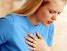
Chest pain when inhaling is not a separate diagnosis, but a symptom that most often indicates irritation of the pleura, pericardium, or chest wall structures. This is why this pain is often stabbing or cutting, and intensifies with deep inhalation, coughing, laughing, turning the torso, or attempting to lie on the affected side. Moreover, the same pain pattern can occur with relatively benign costochondritis, as well as with potentially dangerous conditions, including pulmonary embolism, spontaneous pneumothorax, pleural infection, and pericarditis. [1]

The key clinical idea is that the pleuritic nature of the pain reduces the likelihood of classic ischemic pain, but does not negate a primary assessment for life-threatening conditions. Current chest pain guidelines recommend not attempting to guess the diagnosis based solely on the patient's symptoms, but rather beginning with an assessment of vital signs, an electrocardiogram, markers of myocardial damage, and clinical risk. This approach is necessary precisely to avoid missing a dangerous cause in a patient whose pain initially appears to be "muscle" or "cold-related." [2]

A particular difficulty is that dangerous causes are sometimes masked. For example, pericarditis often causes typical pain that intensifies with inhalation and when lying down, while pulmonary embolism often begins with sudden shortness of breath and stabbing pain without a high fever or severe cough. Therefore, an article on pain when inhaling should focus not on a single organ, but on proper risk triage. [3]

Table 1. The most important causes of chest pain when inhaling and their typical landmarks. [4]

Cause How does pain usually feel? What often accompanies How urgent is it?
Pulmonary embolism Sudden, stabbing, intensifies on inhalation Shortness of breath, tachycardia, sometimes hemoptysis, fainting Urgently, often urgently
Spontaneous pneumothorax Sudden sharp pain on one side Shortness of breath, feeling of lack of air Urgently
Pneumonia, pleurisy, pleural effusion Stabbing pain due to inflammation Fever, cough, weakness Urgently if the condition worsens, otherwise see a doctor quickly
Pericarditis Acute pleuritic pain It gets worse when lying down, and gets better when sitting and leaning forward, and there may be a fever. Urgently
Costochondritis, chest wall pain Local pain Played when pressed or moved Usually there is no urgency after the danger has been ruled out
Acute aortic syndrome Sudden, very severe pain in the chest or back Weakness, sweating, neurological symptoms, drop in blood pressure Immediately

When a symptom is dangerous

The most worrisome combinations include sudden pain on inspiration, dyspnea, hemoptysis, syncope, severe weakness, cyanosis, a drop in blood pressure, or confusion. This combination of signs requires urgent evaluation for pulmonary embolism, pneumothorax, acute coronary syndrome, pericardial tamponade, or acute aortic syndrome. According to current guidelines, hemodynamic instability and respiratory failure determine the level of risk more strongly than a verbal description of pain. [5]

Particularly suggestive of pulmonary embolism are sudden onset, shortness of breath, rapid pulse, recent surgery, prolonged immobility, air travel, pregnancy and the postpartum period, active cancer, estrogen use, and a history of thrombosis. The new 2026 guidelines emphasize that symptoms are often nonspecific, but pleuritic pain and shortness of breath remain the most common complaints, and further decision-making is based on pre-test probability, D-dimer testing, and imaging. [6]

Sudden unilateral pain and shortness of breath, occurring almost immediately, sometimes during complete rest, most often indicate a spontaneous pneumothorax. The British Thoracic Society recommends focusing primarily on the patient's symptoms and physiological stability, rather than solely on the size of the air on the X-ray. Therefore, even with a small pneumothorax, significant shortness of breath makes the situation clinically significant. [7]

Fever, cough, chills, and pain that worsens with a respiratory infection often raise suspicion of pneumonia, pleurisy, or complicated parapneumonic effusion. New guidelines from the American Thoracic Society emphasize the importance of assessing the severity of pneumonia in adults with community-acquired pneumonia, promptly initiating antibiotic therapy, and monitoring for pleural complications, as effusion and empyema often turn "common pneumonia" into a serious problem. [8]

Table 2. Red flags and correct action. [9]

Red flag What to think about first What to do
Sudden pain plus shortness of breath Pulmonary embolism, pneumothorax Urgent care on the same day, in severe cases - ambulance
Pain plus fainting, drop in pressure High-risk thromboembolism, aortic syndrome, tamponade Call an ambulance immediately
Very severe tearing pain in the chest or back Acute aortic syndrome Call an ambulance immediately
Fever, cough, increasing pain Pneumonia, pleurisy, empyema Urgent in-person examination
The pain intensifies when lying down and is relieved when sitting forward. Pericarditis Urgent in-person examination
Local pain is reproduced by palpation Chest wall, costochondritis After excluding the dangerous one - outpatient

Main reasons

Pulmonary embolism

Pulmonary embolism is one of the most common causes of sudden pain when inhaling. The pain is caused by infarction of a portion of the lung or irritation of the pleura, so it is often sharp and stabbing. Unlike many other causes, here the symptoms are often accompanied not only by pain, but also by shortness of breath, increased heart rate, anxiety, and sometimes hemoptysis. [10]

The current diagnostic pathway is structured as follows: first, clinical probability is assessed, then, if the probability is low or intermediate, D-dimer is measured, and if it is elevated, CT scanning of the pulmonary vessels is performed. If the probability is high, imaging is performed without delay. This principle was maintained in European guidelines and was confirmed in the new US guidelines of 2026. [11]

Treatment tactics depend on risk. In stable patients, anticoagulation remains the mainstay of treatment, while some low-risk patients with good social conditions may benefit from early discharge or outpatient management. In cases of hemodynamic instability, systemic thrombolysis, catheter-based therapies, or surgical intervention are considered. [12]

Spontaneous pneumothorax

Spontaneous pneumothorax occurs when air enters the pleural space, causing the lung to partially or significantly collapse. The classic presentation is sudden, unilateral chest pain and rapid onset of shortness of breath. Symptoms often have a greater influence on treatment decisions than the shear size of the pneumothorax on imaging alone. [13]

The British Thoracic Society's 2023 guidelines recommend conservative observation in adults with primary spontaneous pneumothorax if symptoms are minimal and there is no physiological instability. For some patients, an outpatient approach is possible, but only if there is organized follow-up and the ability to quickly reassess the condition. [14]

If pain and dyspnea are severe, the patient is unstable, or there is a pneumothorax secondary to chronic lung disease, aspiration or drainage of the pleural cavity is often required. Recurrent episodes and persistent air leaks often prompt consideration of surgical prophylaxis. [15]

Pneumonia, pleurisy and pleural infection

When the inflammatory process in the lung is located near the pleura, pain upon inhalation becomes very characteristic. Therefore, lower lobe pneumonia, pleurisy, and parapneumonic effusion are often felt as a "prick" or "knife" when taking a deep breath. If the process progresses, fluid and then pus may accumulate in the pleural cavity. [16]

Fever, cough, weakness, sweating, and sometimes shortness of breath are important clinical features. The American Thoracic Society's new guidelines for community-acquired pneumonia emphasize that severity assessment is essential from the outset, as it determines the scope of testing, treatment location, and follow-up time. [17]

If complicated effusion or empyema develops, antibiotics alone are often insufficient. British Thoracic Society guidelines recommend pleural ultrasound, pleural fluid analysis, fine-tube drainage if necessary, and, if residual infection persists, a combination of tissue plasminogen activator and deoxyribonuclease. [18]

Pericarditis

Pericarditis is an inflammation of the sac surrounding the heart. The pain is often very similar to pleuritic pain: it is sharp, intensifies with inhalation, coughing, and lying down, and subsides when sitting and leaning forward. It is this positional component that makes pericarditis one of the most important "cardiac masks" of pain when inhaling. [19]

Current guidelines (2025) consider the diagnosis of pericarditis to be a combination of characteristic clinical features and additional signs, such as a pericardial friction rub, electrocardiogram changes, elevated inflammatory markers, effusion, or signs of inflammation on imaging. This is important because simply "stabbing pain upon inspiration" does not necessarily prove pericarditis and requires confirmation. [20]

Nonsteroidal anti-inflammatory drugs or aspirin plus colchicine remain first-line treatments, along with temporary exercise restriction. The 2025 guidelines specifically emphasize that, in the case of a recurrent inflammatory phenotype, drugs that block interleukin 1 are increasingly important, while glucocorticosteroids are not considered the preferred initial treatment option without specific indications. [21]

Chest wall pain and costochondritis

After ruling out dangerous cardiac and pulmonary causes, localized, reproducible pain is often associated with the chest wall. The most typical variant is costochondritis, when the pain is concentrated near the sternum and intensifies with movement, deep inspiration, and palpation of a specific point. Unlike thromboembolism or pneumothorax, there is usually no pronounced shortness of breath, a drop in blood pressure, hemoptysis, or progressive hypoxemia. [22]

The diagnosis is clinical in most cases. Reviews of ambulatory chest pain emphasize that the reproduction of pain with pressure increases the likelihood of a chest wall source, but should not be the sole reason for omitting a basic evaluation for dangerous causes if there are warning signs or risk factors. [23]

Treatment is usually conservative: temporary limitation of the provoking load, gentle exercise, local heat, a short course of pain medication as prescribed by a doctor, and a gradual return to normal activity. If the pain persists, is atypically localized, or is accompanied by systemic symptoms, a reassessment of the diagnosis is necessary. [24]

Acute aortic syndrome

Although pain from aortic dissection is not always described as "pain on inspiration," this diagnosis should be considered in cases of sudden, severe chest or back pain. Guidelines from the American Heart Association and the American College of Cardiology emphasize that suspicion is heightened by sudden onset, maximum intensity within minutes, a tearing or ripping character, radiating pain to the back, pressure differences between the arms, neurologic symptoms, hypotension, or shock. [25]

This is a rare, but one of the most dangerous causes of chest pain. The patient doesn't need a "home symptom checker"—they need immediate emergency evaluation with imaging and the involvement of vascular or cardiac specialists. This is why sudden, catastrophic pain is always considered an emergency, even if it worsens with breathing and initially appears pleuritic. [26]

Table 3. What helps to distinguish the causes already at the stage of questioning and examination. [27]

Sign Speaks more for
Sudden onset plus shortness of breath Pulmonary embolism, pneumothorax
Fever plus cough Pneumonia, pleurisy, empyema
Worse lying down, better sitting forward Pericarditis
Pain can be accurately demonstrated with a finger and reproduced by pressure Costochondritis, musculoskeletal cause
Very severe pain in the chest or back from the first minutes Acute aortic syndrome
Recent surgery, prolonged immobility, active cancer Pulmonary embolism

Diagnostics

The initial examination begins not with "guessing the diagnosis," but with assessing the severity of the condition. The physician measures blood oxygen saturation, blood pressure, respiratory rate, and pulse rate, assesses consciousness, the presence of cyanosis, asymmetry of respiratory movements, a pericardial friction rub, and localized chest wall tenderness. With any new or worsening chest pain, an electrocardiogram and laboratory evaluation for acute myocardial injury are important. [28]

If symptoms and risk factors suggest pulmonary embolism, clinical probability and D-dimer testing are used. The new 2026 guidelines emphasize that, with low or intermediate probability, a normal D-dimer makes thromboembolism unlikely, while with elevated D-dimer or high probability, a CT scan of the pulmonary vessels is necessary. If contrast imaging is not possible, ventilation-perfusion scanning is an alternative. [29]

If pneumothorax, pneumonia, or effusion is suspected, the initial evaluation is usually a chest X-ray and, if available, a pleural ultrasound. The British Thoracic Society emphasizes that ultrasound is particularly useful for confirming and safely guiding pleural procedures and, in the case of pleural infection, allows for more accurate drainage planning. [30]

If pericarditis is suspected, an electrocardiogram, echocardiography, and inflammatory markers are mandatory. Current guidelines from 2025 expanded the role of advanced imaging, particularly cardiac magnetic resonance imaging, when the diagnosis is unclear, the course is atypical, or the severity of inflammation needs to be assessed. [31]

If the pain is localized and reproducible with palpation, vital signs are normal, and there are no red flags, after a basic cardiopulmonary assessment, the source is most likely in the chest wall. However, even in this situation, the physician considers age, cardiovascular risk factors, the presence of trauma, cough, rash, autoimmune manifestations, and the duration of symptoms to avoid missing a rib fracture, herpes zoster, or a systemic disease. [32]

Table 4. Examinations for chest pain on inspiration. [33]

Study Why is it necessary? When is it especially useful?
Electrocardiogram Exclude acute coronary syndrome, look for signs of pericarditis Almost everyone with new chest pain
High-sensitivity troponin Assess myocardial damage For acute chest pain
D-dimer Exclusion of thromboembolism at low or intermediate probability If thromboembolism is suspected
Chest X-ray Detect pneumonia, pneumothorax, effusion For respiratory symptoms and inflammation
Ultrasound of the pleura Confirm effusion, establish drainage If pleural fluid is suspected
Computed tomography of the pulmonary vessels Confirm or exclude thromboembolism In case of high suspicion or positive D-dimer
Echocardiography Assess the pericardium, effusion, and hemodynamics If pericarditis or instability is suspected
Computed tomography of the aorta Confirm aortic syndrome For sudden tearing pain

Treatment

Treatment for pain when inhaling always depends on the cause, not the pain itself. That's why there's no one-size-fits-all "pain when inhaling" pill. First, the doctor determines whether there's a condition in which pain relief without diagnosis will only delay treatment. This principle is especially important when thromboembolism, pneumothorax, aortic syndrome, and pericarditis are suspected. [34]

In pulmonary embolism, anticoagulation is the mainstay of treatment, and direct oral anticoagulants are often preferred in suitable patients. In severe, high-risk cases with hypotension or shock, systemic thrombolysis is considered, and if contraindicated or ineffective, catheter-based or surgical methods are considered. The new 2026 guidelines specifically link treatment choice to the clinical severity category. [35]

For spontaneous pneumothorax, three main strategies are possible: observation, outpatient management with an air evacuation device, or pleural drainage. The decision depends on symptoms, stability, the presence of chronic lung disease, and the risk of recurrence. Minimally symptomatic primary pneumothorax does not always require immediate drainage, but severe dyspnea almost always changes the approach to active intervention. [36]

In pneumonia and pleurisy, the basis remains timely antibacterial therapy based on the clinical scenario and severity, and in complicated effusions or empyema, a combination of antibiotics and drainage. Guidelines for pleural infection emphasize that in cases of residual infection after drainage, a combination of tissue plasminogen activator and deoxyribonuclease should be considered, rather than single agents. [37]

For pericarditis, first-line therapy includes aspirin or a nonsteroidal anti-inflammatory drug (NSAID) along with colchicine, as well as exercise restriction until clinical resolution. In cases of relapse and inflammatory phenotype, current guidelines from 2025 have strengthened the position of interleukin-1 blocking agents as a preferred escalation over early initiation of glucocorticosteroids. [38]

If costochondritis or other chest wall pain is confirmed after ruling out dangerous causes, a gentle regimen, local measures, and short-term symptomatic treatment are usually effective. However, even here, it is important not to delay re-evaluation if the pain intensifies, shortness of breath, fever, nocturnal symptoms, weight loss, or the lack of response to conservative treatment develops. [39]

Table 5. Treatment depending on the cause. [40]

Cause Basic tactics
Pulmonary embolism Anticoagulation, in case of high risk - reperfusion methods
Spontaneous pneumothorax Observation, ambulatory air evacuation or drainage
Pneumonia Antibacterial therapy, control of complications
Pleural infection Antibiotics plus drainage, sometimes intrapleural enzyme therapy
Pericarditis Anti-inflammatory therapy plus colchicine, exercise restriction
Costochondritis Symptomatic treatment and reduction of the provoking load

What you can and can't do before seeing a doctor

If the pain is new, intense, appears suddenly, or is accompanied by shortness of breath, don't wait for it to "go away on its own." Don't rely solely on the fact that the pain is stabbing or dependent on inhalation: this doesn't automatically make the situation safe. In scenarios involving shortness of breath, fainting, severe weakness, hemoptysis, or catastrophic chest and back pain, there's only one correct course of action: seek emergency medical attention. [41]

Self-medication with painkillers is only permissible for mild pain without red flags and without attempting to delay an in-person examination for several days if the cause is unclear. It is especially dangerous to "kill" the pain and continue physical activity if pericarditis is possible, or to ignore sudden shortness of breath if thromboembolism or pneumothorax is likely. [42]

Before the examination, it is helpful to record the time of pain onset, its relationship with breathing, coughing, body position, physical activity, chest compression, as well as the presence of fever, shortness of breath, recent travel, surgery, trauma, immobilization, pregnancy, cancer history, and previous thrombosis. This information directly influences the choice of examinations. [43]

Table 6. When one can think about outpatient tactics and when not. [44]

Situation Outpatient treatment is possible Outpatient treatment is not allowed.
Confirmed chest wall pain without red flags Yes No, if the pain increases or systemic symptoms appear
Suspected primary small pneumothorax Sometimes, only after an in-person assessment and organized observation No, if there is severe shortness of breath or instability
Confirmed low-risk thromboembolism Sometimes in selected patients No, if there is high risk or instability
Pneumonia with pleural pain Sometimes with mild course of the disease No, if there is respiratory failure, effusion, septic signs
Pericarditis Sometimes, if there are no high risk factors No, if there is effusion, instability, myopericarditis, severe course

Prognosis and prevention

The prognosis is entirely determined by the underlying cause. For musculoskeletal pain and costochondritis, it is usually favorable. For pericarditis, the outcome is also generally good, but relapses are possible, making colchicine and observation particularly important. For pneumonia, the prognosis depends on age, comorbidities, and the presence of pleural complications. For pulmonary embolism and aortic syndrome, the outcome directly depends on the speed of diagnosis and treatment. [45]

Prevention is based on the underlying cause: quitting smoking reduces the risk of recurrent spontaneous pneumothorax, preventing venous thromboembolism is important after surgery and periods of immobility, vaccination and chronic disease control reduce the risk of severe pneumonia, and prudent management of exercise and inflammatory diseases helps reduce the risk of pericardial recurrence. There is no universal prevention for "inspiratory pain" itself, as it is not an independent nosology. [46]

FAQ

1. Can chest pain when inhaling simply be from a muscle?
Yes, it can. If the pain is localized, reproduced by palpation or movement, and is not accompanied by shortness of breath, fever, fainting, or other red flags, the source is likely in the chest wall. But it's safe to definitively consider it "muscular" only after ruling out dangerous causes. [47]

2. Does worsening pain on inhalation mean it's not the heart?
No. This type of pain is indeed more typical of the pleura, pericardium, or chest wall, but pericarditis is a cardiac cause, and some dangerous conditions cannot be ruled out based on the pain description alone. Therefore, when new chest pain occurs, a clinical algorithm is needed, not guesswork based on sensation. [48]

3. When should you call an ambulance immediately?
Immediately – if the pain is sudden and severe, accompanied by shortness of breath, fainting, severe weakness, cyanosis, a drop in blood pressure, hemoptysis, or is tearing and radiating to the back. These are scenarios that require consideration of thromboembolism, pneumothorax, acute aortic syndrome, and other emergency conditions. [49]

4. What is the most important test if pulmonary embolism is suspected?
Not just one test, but the correct sequence: first, clinical probability, then D-dimer if the probability is low or intermediate, and then CT scan of the pulmonary vessels if indicated. An elevated D-dimer alone does not establish a diagnosis, and a normal result is useful only in the correct clinical context. [50]

5. Why is pericarditis pain relieved when sitting and leaning forward?
Because this position reduces mechanical irritation of the inflamed pericardial sac. This is one of the classic signs of pericarditis, although not absolute: the diagnosis still requires confirmation with additional data. [51]

6. Can I treat this type of pain at home with antibiotics at random?
No. If the cause isn't bacterial pneumonia, antibiotics won't help. But if it's a pleural infection, pneumothorax, pericarditis, or thromboembolism, wasting time is dangerous. Therefore, treatment should be initiated only after an in-person assessment and confirmation of the probable cause. [52]

7. Can a normal chest x-ray immediately exclude all dangerous causes?
No. Chest x-rays are useful for detecting pneumonia, effusions, and some pneumothoraces, but they do not exclude pulmonary embolism and are not a definitive test for aortic syndrome or pericarditis. Therefore, the choice of imaging depends on the clinical scenario. [53]

8. Can pain persist for weeks after a musculoskeletal cause has been confirmed?
Yes, it can, especially if coughing, chest strain, or abnormal trunk movements continue. However, if pain increases, fever, shortness of breath develops, or the expected improvement fails, the diagnosis should be reconsidered. [54]

Key points from experts

  • Chest pain when inhaling is a symptom, not a diagnosis, so the key is not to describe the pain, but to triage the risk and find the cause. [55]

  • Sudden pleuritic pain plus dyspnea is one of the most important scenarios to exclude pulmonary embolism and pneumothorax.[56]

  • Pericarditis remains an important cardiac cause of pain on inspiration; typical relief from sitting and leaning forward is a helpful clue but not a diagnosis in itself.[57]

  • In case of pleural infection, not only antibiotics are used, but also timely drainage with ultrasound guidance and, if necessary, intrapleural enzyme therapy. [58]

  • Local reproducible pain does indeed more often point to the chest wall, but this does not excuse a basic assessment for dangerous causes when red flags are present.[59]

  • Any sudden, very severe chest or back pain within the first few minutes should be considered as potential acute aortic syndrome until proven otherwise.[60]