Medical expert of the article

New publications
Dental exostosis
Last reviewed: 29.06.2025

All iLive content is medically reviewed or fact checked to ensure as much factual accuracy as possible.
We have strict sourcing guidelines and only link to reputable media sites, academic research institutions and, whenever possible, medically peer reviewed studies. Note that the numbers in parentheses ([1], [2], etc.) are clickable links to these studies.
If you feel that any of our content is inaccurate, out-of-date, or otherwise questionable, please select it and press Ctrl + Enter.
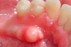
In dentistry, there is such a term as "tooth exostosis". It is a bone overgrowth that has the appearance of a protrusion in the area of the gum or jaw. As a rule, there is no pain. The main problem is the gradual increase in overgrowth, which leads to discomfort in the oral cavity, an increased risk of damage to the gum tissue, the occurrence of speech and digestive disorders. Tooth exostosis often interferes with the use of dentures and implants, contributes to the formation of an incorrect bite and chin asymmetry. The initial stage of formation of the neoplasm is imperceptible, and as it grows, the patient can easily feel the pathological growth with the tongue. [1]
Causes of the tooth exostosis
A dental exostosis is a bony protrusion that appears in the maxillary or mandibular dentition, most often after tooth extraction, traumatic injury, or due to an age-related process in the alveolar ridge. Most commonly, maxillary exostoses form from the cheek portion of the alveolar process. Mandibular exostoses are found mainly on the lingual part of the premolars or molars, incisors, canines. [2]
Symmetrical overgrowths at the sites of the lower small molars are found in people with adentia.
Tooth exostosis is an acquired condition. The most common causes of its occurrence are as follows:
- Tooth extraction, accompanied by an incomplete process of smoothing of the well margins;
- Dental extirpation with severe tissue damage;
- Injuries, jaw fractures, improper bone fusion;
- Dysplastic processes in the jaw.
Risk factors
Hereditary factors play an important role in the appearance of dental exostosis. Some patients have a genetic predisposition associated with a congenital tendency to bone abnormalities.
Bone overgrowth of a tooth can occur at any age, but it is most often detected in adolescents - at the stage of intensive bone growth. In infants and preschoolers, the problem is much less common.
Among the other most likely preconditions for the development of pathology:
- Endocrine disorders;
- Infectious inflammatory processes;
- Dental malocclusions;
- Hypercalcemia;
- General poor dental health.
Pathogenesis
Specialists do not have an absolute understanding of the pathogenetic processes of dental exostosis. It is known that:
- The risks of the problem increase with the development of inflammatory or tumor changes in the bone tissue;
- Pathological growth can be a consequence of tooth extraction, which is especially relevant in the case of problematic removal of retined or dystopian wisdom teeth and is associated with increased traumatization;
- The formation of tooth exostosis can occur against the background of prolonged or long-standing periodontal disease;
- Outgrowths often occur after tooth extraction without smoothing of the lunar margins;
- Bony prominences may result from jaw trauma, inadequate ratio of damaged jaw elements, or old fractures;
- Exostoses of osteogenic dysplastic etiology sometimes develop in the periphery.
Symptoms of the tooth exostosis
In the vast majority of cases, tooth exostoses are not accompanied by any obvious symptoms. The problem is detected during a dental appointment or in the preparatory phase before prosthetics.
Patients most often experience no discomfort when opening the mouth or moving the jaw. The mucosa over the exostosis is pale pinkish, without obvious pathological signs, not adherent to the bone tissue.
As exostoses grow, the mucosa may thin, and then there is an increased likelihood of its damage, injury by food particles and teeth. When palpating the area of the growth, a dense protrusion with a smooth or bumpy surface is detected, painless. [3]
Nearby lymph nodes are not enlarged, the general well-being of patients does not suffer.
Possible additional symptoms include:
- Changes in the shape of the gum, the jaw;
- Jaw asymmetry;
- Swelling of the gum, swelling of the surrounding tissues;
- Pain associated with pressure on the teeth and surrounding tissues.
What does an exostosis look like after a tooth extraction?
Exostosis after tooth extraction is not uncommon, as well as after other jaw traumas and injuries. In this situation, intense bone growth is due to a specific evolutionary defense mechanism involving the repair of damaged tissue. Such overgrowth usually needs to be removed. [4]
Often exostosis on the gum after tooth extraction is formed in the area of extracted molars (VI, VII, VIII teeth), which play an important role in the processes of primary food processing. Exostosis after wisdom tooth extraction is even more common.
Symptomatology of the appearance of growths is rather scarce. Often the problem is detected by a dentist during a preventive or therapeutic examination.
Exostosis of the jaw after tooth extraction may present with the following signs:
- Sensation with the tongue of a dense mass with a smooth or rough surface;
- The feeling of a foreign object in the mouth;
- When localized in the temporomandibular joint - jaw dysfunction;
- Pallor of the mucosa in the area of the growth.
If there are no serious disorders of the temporomandibular joint, exostosis after tooth implantation does not cause problems with opening and closing the mouth. The neoplasm is not caused by infectious diseases and is usually not accompanied by fever or purulent discharge, but such signs may indicate the development of complications.
Complications and consequences
A tooth exostosis should preferably be removed as soon as it is detected. Even a small and harmless looking growth in most cases has a tendency to grow further. This is associated with the likelihood of complications.
Exostoses can:
- To grow to a rather large size;
- Damage adjacent tissues, adversely affect the growth and localization of adjacent teeth; [5]
- Make it more difficult to perform oral hygiene procedures;
- Create problems for a proper bite;
- Swelling, inflammation, infection;
- Interfere with dental treatment and prosthetics.
A large tooth exostosis often prevents you from correctly pronouncing letters, words, and chewing food.
Transformation of this bone mass into a malignant tumor was not observed.
Diagnostics of the tooth exostosis
Diagnostic measures are performed by a dental surgeon. They include interview and clinical examination of the patient, radiographic examination. The main task of the dentist is to identify the problem and exclude possible other pathological processes. Thus, differential diagnosis is carried out to exclude:
- The appearance of a dental protrusion (additional occlusal cusps);
- Dental anomalies (dilated odontomas);
- Adamantine;
- Abscesses, root cysts;
- Gingival recessions;
- Gingival cysts, gigantocellular or fibrous epulis, pyogenic granuloma;
- Of dental anomalies;
- Other cystic neoplasms and jaw abnormalities;
- Of multiple maxillary exostosis. [6]
During the examination, the specialist can determine the presence of a dense protrusion without adhesion with adjacent tissues. Stale exostoses may have lesions on their surface, ulcers. In advanced cases, stomatitis may develop.
On radiograph, a characteristic bony neoplasm with clear configurations and no destructive bony changes can be identified.
In complex cases, to clarify the diagnosis may be appointed computer or magnetic resonance imaging, biopsy.
What do need to examine?
Who to contact?
Treatment of the tooth exostosis
It is impossible to get rid of tooth exostosis on your own: the overgrowth is surgically removed. The operation is performed by a dental surgeon. Among the likely contraindications to intervention:
- Diabetes;
- Disorders of the endocrine apparatus and adrenal glands;
- Clotting disorders.
If the neoplasm is small (up to 2-3 mm) and the patient does not complain of discomfort, surgical treatment is postponed and dynamic observation is prescribed instead. If the growth increases, presses on the tongue, cheek, neighboring teeth, if it interferes with dental prosthetics or treatment, then surgical correction becomes mandatory.
Before removing the bone overgrowth, the dentist anesthetizes the surrounding tissue and makes a gingival incision of the required length. Next, he saws off the tooth exostosis, cleans the sharp protrusions, after which he resurfaces the soft tissues and sutures the wound. The duration of manipulation can vary from 60 to 120 minutes, which depends on the size of the formation and the availability of access to it.
During the first two to three days after the intervention, it is necessary to take care of the wound, rinse it with antiseptic solutions (as prescribed by the doctor), observe oral hygiene. It is important for a while to exclude from the diet rough, hard, hot, too acidic and spicy food. It is also not allowed to take alcoholic drinks and smoking.
During the recovery period, you should not engage in active sports, perform deep bending and jumping. It is advisable to avoid stressful situations, take more rest and get a good night's sleep.
Swelling and pain may be bothersome at first. To alleviate the condition, you should consult a doctor who will prescribe appropriate analgesics and non-steroidal anti-inflammatory drugs. It is necessary to eat only liquid food, drink water, use dairy products, rinse the mouth regularly to avoid infection and the development of purulent process in the wound.
With a qualified approach to surgical treatment and patient compliance with all medical recommendations, recovery is fast and there are no complications. It is necessary to visit a doctor if there is severe and prolonged pain, fever, swelling worsens.
Prevention
As part of preventive measures, it is important to visit doctors in a timely manner, provide treatment for any dental pathologies, and regularly visit the dentist for routine checkups. Among additional recommendations of specialists can be distinguished such as:
- Quality brushing twice a day;
- The use of dental floss, special mouthwashes;
- Eating enough plant foods and dairy products;
- Avoiding trauma to the jaws, teeth, and oral tissues;
- Routine visits to the dentist at least twice a year.
Attention to yourself and your health is an important component of the prevention of any disease. It is much easier to prevent the development of a disease than to put all your efforts into treating it later.
Forecast
The prognosis can be classified as favorable. After exposure to etiologic factors and surgical removal of the pathologic growth, the probability of recurrence is virtually nil.
Independent attempts to get rid of the problem are always unsuccessful and even dangerous. This is due to the fact that we are talking about a bone growth, which is quite dense in its structure. If you try to remove it yourself, it will irreversibly lead to trauma to the soft tissues and the development of an infectious process. To avoid additional troubles, removal of the neoplasm should be carried out by a dental surgeon.
So, why you can't remove a tooth exostosis on your own:
- It traumatizes the gum and the jaw itself;
- It could cause the infection to spread;
- It will complicate further diagnostic and therapeutic measures.