Medical expert of the article

New publications
Gingival exostosis
Last reviewed: 29.06.2025

All iLive content is medically reviewed or fact checked to ensure as much factual accuracy as possible.
We have strict sourcing guidelines and only link to reputable media sites, academic research institutions and, whenever possible, medically peer reviewed studies. Note that the numbers in parentheses ([1], [2], etc.) are clickable links to these studies.
If you feel that any of our content is inaccurate, out-of-date, or otherwise questionable, please select it and press Ctrl + Enter.
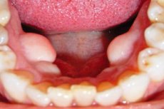
Pathologic bone growths are not only found in orthopedics, but also in dentistry. One of the varieties of such a problem is a gingival exostosis, which is also called a bone spike. This neoplasm is formed from periodontal cartilage and belongs to a number of benign tumors with no tendency to malignancy. Despite the apparent "harmlessness" of exostosis, it causes significant discomfort to the patient, worsens the functions of speech and chewing food, and in general negatively affects the quality of life. [1]
Causes of the gingival exostosis
Gingival exostosis is a pathology that can occur in a person of any age and gender. A certain role is played by genetic predisposition: hereditary exostoses are more often formed in childhood, with their growth intensifying during the period of hormonal restructuring - in particular, at the stage of puberty.
Experts point out the following most common reasons for the formation of gingival exostosis:
- Purulent foci in the periodontium, fistulas and flux, atrophic and destructive processes in the bone;
- Developmental defects of a particular tooth;
- Chronic course of periostitis;
- Hormonal changes that affect bone structure;
- Syphilitic bone lesions;
- Improper or hyper-traumatic extraction of a tooth;
- Injuries to the jaw, including complete or partial fractures, dislocations.
In some children, gingival exostosis forms during the replacement of baby teeth with molars.
Statistically, the most common causes of gingival exostosis are:
- Complications of tooth extraction;
- Jaw injuries accompanied by active regeneration of the affected tissues with active cell division and overgrowth.
Often, exostosis forms in patients who refuse chin splint fixation and fail to provide immobility of the jaw during the period of bony recovery after fracture. [2]
Risk factors
The provoking factors for the occurrence of gingival exostosis are said to be:
- Hereditary predisposition; [3]
- Traumatic injuries, both directly to the gum and to the jaws;
- Bite disorders and other defects, including congenital defects;
- Acute and chronic pathologies in the oral cavity.
Specialists also highlight other likely factors:
- Metabolic disorders;
- Chronic intoxication;
- Bad habits.
Genetically determined gingival exostosis is more often multiple, its location is usually symmetrical.
Pathogenesis
Bone and cartilaginous growth occurs under the soft tissues of the gum. At an early stage of development, the problem does not make itself known for a long time: at first, the exostosis has the appearance of a cartilaginous neoplasm, which after some time hardens and transforms into a bony protrusion. A dense bone capsule similar to a shell is formed on its surface.
Visually, a gingival exostosis can have different configurations, from oblong or spiky, to rounded or mushroom-shaped. The size can also vary from a few millimeters to 1-2 cm. Exostoses are more often single, less often - multiple, located symmetrically.
Over time, the neoplasm progresses, the growth becomes larger, begins to create problems in chewing food, interferes with normal speech function. In neglected cases, gingival exostosis causes jaw deformation, bite disorders and tooth growth. The defect is visualized with the naked eye, taking the form of a thickening under the gum. [4]
In some patients, bone and cartilage growths grow very slowly and do not cause any trouble for decades. Such exostoses are discovered accidentally, particularly during radiography or routine dental checkups.
Symptoms of the gingival exostosis
At the initial stage of development of gingival exostosis, there are no obvious symptoms. In the area of the gum is just palpable small thickening, which practically does not interfere in any way, is not accompanied by pain. However, after some time, the growth increases. At this stage of development, the first symptoms appear:
- A persistent foreign body sensation in the mouth;
- Redness, enlargement of the gum in the area of the pathologic focus;
- Changes in speech (if the growth is large);
- Sometimes - pain when palpating the neoplasm).
The appearance of a thickening is not associated with tissue infection, does not have a tendency to malignancy. It is associated solely with increasing discomfort, which is reported by almost all patients. In some cases, exostosis adds problems in terms of certain dental manipulations - for example, dentures.
Exostosis on the gum after tooth extraction can form at the base of the incisors or canines. The pathologic formation has a lump-shaped or pyknotic form.
The growth begins its formation asymptomatically. At the first stage, a small, slowly increasing mass appears under the gingiva, which can be detected only accidentally. As it grows, the corresponding symptoms appear:
- A visualized "bump" that is hard when felt;
- Lightening of the gingiva in the area of the pathological focus;
- Constant feeling of the presence of a foreign object in the oral cavity;
- Chewing and speech problems;
- In advanced cases - jaw deformity, facial asymmetry.
Gingival exostosis can be accompanied by pain only in the close location of nerve fibers and endings, when the growth presses on the root of the tooth, or when the inflammatory process develops. Inflammation may be caused by systematic friction of the soft tissues of the lips or cheek on the pathological growth with the penetration of an infectious agent into the formed wound. In such a situation, the neoplasm swells, reddens, there is an unpleasant odor from the oral cavity. [5]
Complications and consequences
Leaving gingival exostosis untreated is not recommended, since such neoplasms tend to increase constantly. In most cases, surgical removal of the pathological overgrowth is used: it is the only effective way to completely eliminate the defect.
If the problem is not corrected, it can negatively affect dentures, interfere with normal speech and eating, deform the jaw, and affect the bite of the teeth.
Will exostosis on the gum go away on its own? If its appearance is associated with intoxication, hormonal or metabolic disorders in the body, which can be eliminated, then small-sized growths (up to 2-3 mm) can regress. However, in most cases it is necessary to seek help from a surgeon.
It is important to know that in some patients, gingival exostosis is able to recur, which is especially relevant for those who have a genetic predisposition to this pathology.
Gingival exostosis refers to benign growths with no tendency to transform into a malignant process.
Diagnostics of the gingival exostosis
Since this pathology on the gum practically does not manifest itself symptomatically, it is most often detected during a dental examination. Sometimes a suspicious growth is indicated by the patient himself.
After visual inspection and palpation of the pathological formation, the doctor may refer the patient for additional examinations: radiography, orthopantomogram. Based on the results of diagnosis, the specialist establishes a diagnosis, describes the characteristics of exostosis (localization, size, configuration, complications): the growth is usually round or spiky, without adhesion to the gingival tissues. [6]
If necessary, the doctor will prescribe additional tests:
- Biopsy with histologic analysis;
- A CT or MRI;
- Laboratory tests (general and biochemical blood tests, Wasserman reaction).
Differential diagnosis
In some cases, bone growths of the gingiva reach large sizes, acquire an atypical configuration, without being accompanied by pain syndrome. In such situations, it is very important to make a differential diagnosis - in particular, from cystic formations, epulis, mesenchymal tumors (osteochondroma). For this purpose, the doctor prescribes additional examinations for the patient:
- Computer tomography to clarify the size and localization of the neoplasm, its location in relation to the dental roots and other dentoalveolar components;
- Biopsy to rule out malignancy.
If indicated, it is possible appointment of magnetic resonance imaging, consultation of orthodontist, oncologist.
The differential diagnosis is often able to identify:
- Root fractures, purulent foci;
- Cracks and other bone injuries;
- Hidden formations of other exostoses.
Who to contact?
Treatment of the gingival exostosis
Treatment of gingival exostosis without surgery is possible only if the appearance of the problem is associated with disorders that can be eliminated conservatively. For example, if the root cause of the formation of the growth was metabolic disorders, and the size of the neoplasm is within 3 mm, the therapy of the underlying disease and correction of metabolism is prescribed. With the normalization of the state of the body, such exostoses may well regress. [7]
In situations where the cause of the growth could not be established, or if it is not possible to influence this cause, surgical treatment is prescribed, which consists of surgical removal of the gingival exostosis. Surgery is strongly recommended:
- If the neoplasm is rapidly enlarging;
- If there's any pain;
- If there is facial asymmetry, bite abnormalities;
- If there are problems with speech and eating;
- If gingival exostosis prevents dental implants or dentures from being performed.
Surgery may be refused if the patient is found to have:
- Clotting disorder;
- Diabetes;
- Pronounced hormonal disorders that prevent further wound healing;
- Malignant neoplasms.
The standard surgical manipulation for the removal of gingival exostosis is performed as follows:
- An anesthetic is injected into the gum, the oral cavity is treated with an antiseptic agent;
- Perform a gingival incision, expose the area of pathologic neoplasm;
- The protrusion is removed using a drill with a special attachment, then this place is carefully cleaned;
- If bone damage is detected, the defect is covered with a special plate;
- The removed tissue is put back in place and sutured.
Often practiced and so-called laser therapy: after treating the area of the pathological focus on it is directed laser beam, which heats and "melts" excessive tissue overgrowth. This procedure is characterized by easier and faster tissue recovery time.
Surgery can last from 1 to 2 hours, depending on the complexity of the manipulation and the method used.
What to do after removal of exostosis on the gum? The main rehabilitation stage lasts for about a week, but the full recovery of tissues can be spoken about about 20-30 days after surgery. During this period it is recommended:
- Take analgesics and non-steroidal anti-inflammatory drugs as prescribed by a doctor (the course may be 3-5 days);
- Rinse the mouth with antiseptic solutions to prevent the development of infectious pathologies;
- When indicated, apply topical preparations to stimulate tissue repair and accelerate wound healing;
- In the presence of purulent inflammation take antibacterial agents (as prescribed by a doctor).
During the recovery period, it is important to adhere to the following recommendations:
- Eliminate the consumption of rough, hard, rigid foods;
- Consume only warm, soft foods and beverages;
- Do not touch the wound area with fingers, any objects, or tongue;
- During the first 48 hours after surgery, limit physical activity, avoid sharp bending, do not lift weights;
- Exclude smoking and alcohol intake (cigarette smoke and alcoholic beverages provoke irritation of damaged tissues and worsen the course of recovery processes).
Most patients after the removal of exostosis have painful gingiva, swelling, sometimes the temperature rises to subfebrile. This condition is a normal reaction of the body to tissue damage. As the healing process progresses, the feeling of well-being normalizes.
Prevention
In order to avoid the formation of exostoses, it is necessary to adhere to these medical recommendations:
- Maintain dental and oral hygiene;
- Systematically visit the dentist even if your teeth are in normal condition - for preventive check-ups;
- Seek medical attention in a timely manner for any illnesses, including dental diseases.
Measures should be taken to avoid possible jaw injuries. In particular, athletes should wear protective equipment (helmets, mouth guards, etc.) when practicing boxing, wrestling and other injury-prone sports.
Also do not forget about the possibilities of self-diagnosis: if the first suspicious symptoms appear, it is important not to delay to see a doctor.
Forecast
Benign bone and cartilage overgrowths can occur without a clear root cause. In this case, the only effective method of treatment is considered to be surgery. The intervention is minimally traumatic, in most cases does not require general anesthesia and complex equipment.
If the neoplasm has been removed, but its cause has not been eliminated, there is a certain amount of risk of tissue overgrowth - recurrence, in the same place or with a change in localization.
Self-resorption of exostosis is possible if it appeared in childhood, or after removing the cause of its appearance (for example, after correction of metabolism or normalization of the hormonal background). If the excrescence does not disappear, or even increases, it is advisable to remove it. Choosing a doctor for the operation, it is desirable to base not so much on the cost of intervention, but on the qualifications and experience of the dentist or surgeon. In general, gingival exostosis has a favorable prognosis.