Medical expert of the article
New publications
Exostosis of the big toe
Last reviewed: 29.06.2025

All iLive content is medically reviewed or fact checked to ensure as much factual accuracy as possible.
We have strict sourcing guidelines and only link to reputable media sites, academic research institutions and, whenever possible, medically peer reviewed studies. Note that the numbers in parentheses ([1], [2], etc.) are clickable links to these studies.
If you feel that any of our content is inaccurate, out-of-date, or otherwise questionable, please select it and press Ctrl + Enter.

Exostosis is a not uncommon pathology that is manifested by excessive growth of bone tissue on the surface of the bone. Exostosis of the big toe is most common in the foot. The overgrowth can have a linear, spherical or ridged shape, it can occur in almost any segment of the bone, including under the nail.
Epidemiology
Exostosis, or osteochondroma, is the most common skeletal tumor entity. Bone and cartilage growths account for about 20% of all cases of bone neoplasms and almost 40% of all benign bone tumors. The majority of such pathologies are detected in patients under 20 years of age - and accidentally during radiography, because most often at a young age, the growths develop asymptomatically. Pain only appears as the growths grow when they start to be squeezed by shoes.
In young children, the appearance of exostoma of the big toe can be associated with failure to comply with the rules of prevention of rickets, excessive intake of preparations containing vitamin D.
The problem is most often found in women (about 20-40% more often than in men).
Causes of the exostosis of the big toe
The main cause of this type of exostosis is regular traumatic impact on the big toe area. Traumatization can occur:
- Regular friction due to wearing tight, narrow shoes;
- When walking long distances or running for long periods of time;
- In professional dancing (ballet), cycling;
- For repetitive mechanical trauma to the thumb;
- After surgical removal of the nail plate due to ingrowth;
- When the nail is thinning as a result of mycosis or other pathological processes.
Exostosis of the big toe is often found in obese people, professional athletes, dancers, and those whose professional activity involves increased load on the foot and lower limbs in general. As a result of foot injuries, the load on the big toe increases - mainly during motor activity, walking, running. This contributes to the formation of bone and cartilage growths - exostosis. [1]
The hereditary factor is also of considerable importance. Translocation t(X;6) (q22;q13-14) is reproducibly associated with subfoot exostosis, [2], [3] implying that it is a true neoplasm and not a reactive process in response to trauma. Often, exostoses of the thumb "haunt" relatives of more than one generation.
Risk factors
Exostosis of the big toe in many cases is a hereditary disorder. That is, a person has a predisposition to the appearance of such formations, which is activated under the influence of relevant factors:
- Wearing narrow, tight, uncomfortable shoes;
- Metabolic disorders, endocrine function, obesity;
- Constant intake of hormonal drugs, hormonal disorders in the body;
- Infectious and inflammatory diseases;
- Elevated calcium levels in the body;
- Periosteum developmental defects.
Risk groups include professional athletes (runners, cyclists, soccer players), dancers (ballet), as well as people whose profession involves a long stay "on the feet" and is accompanied by frequent hypothermia or trauma to the extremities.
Pathogenesis
Exostosis of the big toe is an osteochondral tumor of benign character, the appearance of which is caused by traumatic or inflammatory changes in the tissues, especially often - wearing uncomfortable, unsuitable shoes.
Exostosis can form as single (solitary) or multiple growths. A single isolated exostosis of the big toe is rare. Most patients have similar growths on other bony structures, such as the clavicles, spinal column, humerus, femur, and tibia.
The full pathogenetic mechanism of exostosis formation is still unknown and under investigation. Presumably, solitary growths may be the result of displacement of the lamina epiphysis, which, in turn, is explained by failures in embryonic development, irradiation, exposure to ionizing rays. The epiphysis is a cartilaginous tissue localized under the bone head. Epiphyseal cells are constantly mitotically dividing, which provides an increase in the length of human bone as the skeleton grows and develops. After some time, the distal structures of the epiphysis ossify, and bone tissue is formed. If at this stage, under the influence of any provoking factor, part of the epiphysis plate is displaced against the background of further cell division, a new ossification in the form of exostosis is formed. That is, at first it is cartilaginous tissue, which over the years thickens, hardens, with the preservation of the cartilaginous apex. The exostosis of the big toe increases as the overall growth of the bone increases.
Genes are involved in the development of multiple exostosis: the pathology is usually attributed to a number of hereditary diseases. Massive growths affecting not only the big toe, but also other bones of the skeleton, are often detected in childhood. Such a problem requires medical supervision in dynamics, since there is a risk of malignization of such formations. The risk of malignization of a single exostosis of the big toe is relatively low and is less than 1%.
Symptoms of the exostosis of the big toe
In many patients, especially at the initial stage of the disease, exostosis of the big toe does not show any painful symptoms. When it forms on the outer-lateral surface of the thumb bone, there may be signs of soft tissue hyperkeratosis, although a full-fledged callus is not formed. When attempting to remove the skin seal, the sensation of discomfort does not disappear, and the keratinization zone is formed again.
Over time, when the exostosis enlarges, the growth begins to traumatize soft tissues, and chronic joint inflammatory processes develop. From this point on, there is a pronounced discomfort and pain syndrome, especially noticeable when walking in shoes. If you try to palpate the zone of exostosis, then on the big toe you can detect a protruding bone seal with a rough or smooth surface.
During the active growth of the exostosis, the big toe becomes curved, which can manifest itself as a so-called valgus deformity: the toe deviates from its normal axis towards the other toes. As a consequence, the toes nearest to it are also deformed - in particular, they acquire a hammer-shaped configuration. This is a serious aesthetic and physical defect.
There is swelling of the foot and fingers (especially in the afternoon), a feeling of numbness and "creeping goosebumps".
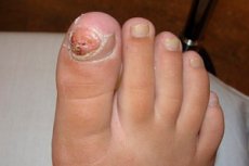
Subnail exostosis is characterized by the appearance of a bulge at the end of the phalanx of the thumb. Visually, the outgrowth resembles a compacted nail roller. Additional symptoms include:
- Pain when walking or pressing on the area of the growth;
- Abnormal growth of the nail plate, detachment or ingrowth of the nail;
- Swelling, redness of the big toe;
- The formation of omosoles.
Complications and consequences
Exostosis of the big toe is prone to progression. It is especially common if there are factors that negatively affect the foot area:
- Overweight;
- Regular carrying/weight lifting;
- Prolonged "on your feet."
- Poor quality or improperly fitting shoes.
- The possibility of malignancy of the bone growth cannot be ruled out.
The risks of recurrence of neoplasm growth remain even after surgical removal. The main way to prevent recurrence is to carefully follow the doctor's recommendations after the intervention:
- Wearing comfortable and good quality shoes;
- Avoiding overloading of the operated finger area;
- Limiting the strain on your legs;
- Weight control;
- Preventing hypothermia of the feet.
If the above rules are followed and lifestyle adjustments are made, the likelihood of recurrence of thumb exostosis is minimized.
Diagnostics of the exostosis of the big toe
If the first signs of exostosis of the big toe appear, it is necessary to visit an orthopedist without delay. Most often, it is not a problem for the specialist to diagnose exostosis during the examination. However, in order to clarify some points, the collection of additional information is required. In particular, the doctor collects data on professional characteristics, lifestyle of the patient, the general condition of the body. The information obtained helps to determine the optimal treatment scheme.
In addition, the specialist specifies the nature of the pain syndrome, localization, duration, signs of neurological disorders, limited physical activity, etc.
As part of the orthopedic examination, the doctor assesses the degree of mobility of the joints, the ability to perform active and passive movements. Additionally, he determines the state of the vascular network, skin of the feet and lower legs, as well as sensitivity and tone of the musculature. These manipulations help to clarify the likely causes of the formation of exostosis and combined pathologies.
This is followed by an instrumental diagnosis:
- Radiography is the main technique used to diagnose exostosis of the big toe. X-rays help to visualize the bones and articulations, and the area of exostosis directly on the image has the appearance of a protruding bony part. It is possible to perform radiography in several projections (2 or 3).
- Ultrasound is a standard procedure that may be ordered to further evaluate tissue conditions.
- Computed tomography can clarify and supplement the information obtained during conventional radiography, as well as determine the internal structure of the exostosis.
- Magnetic resonance imaging will be useful if malignization of a bone-cartilaginous growth is suspected.
Diagnosis is prescribed, depending on the specific situation and the suspected pathology.
Differential diagnosis
During the initial diagnosis, exostosis of the big toe can be mistaken for another pathology. In active stages of development, the growth, accompanied by pain and redness, has many similarities with inflammatory and gouty arthritis. It is important to note that pain due to gout appears abruptly, while pain with exostosis occurs gradually, often after prolonged wearing of shoes. In addition, for differential diagnosis, it is important to determine the uric acid level (this level is increased in patients with gout).
Many forms of arthritis have similarities to exostoses. For example, in septic arthritis, there is swelling and redness.
The possibility of surgical and traumatic arthropathy and valgus curvature of the foot should also be considered.
If there is a history of previous trauma, a dislocation of the thumb, a fracture (including one with malunion) must be distinguished.
Who to contact?
Treatment of the exostosis of the big toe
To relieve pain and eliminate inflammation, the patient is prescribed conservative treatment. It is selected individually, taking into account the severity of exostosis, the general condition of the patient. In most cases, it is appropriate to use external preparations (ointments, creams) based on nonsteroidal anti-inflammatory drugs, as well as similar drugs for oral administration. It is important to understand that such medications will not be able to eliminate exostosis of the thumb, but only help to relieve symptoms.
The only method to completely eliminate the exostosis is surgical treatment, which is indicated:
- For large exostoses;
- An obvious deformity of the thumb;
- Persistent pain syndrome;
- The occurrence of complications (including malignancy).
The intervention is technically uncomplicated and can be performed using local anesthesia. In most cases, the technique of marginal resection of the growth is used. A transverse incision is made in the area of projection of the neoplasm. The length of the incision depends on the size of the exostosis and is most often a few millimeters. The soft tissue is carefully separated from the bone for better visualization of the neoplasm and determination of its boundaries.
Using surgical instruments, the doctor carefully removes the bone mass within the unchanged tissue. The entire overgrowth together with the cartilaginous tip must be removed. If this is not done, the problem may recur after a while. The operation is completed by actively washing the wound with physiological and antiseptic solution, suturing and applying a sterile dressing.
If, in addition to exostosis, there is a curvature of the phalanx of the big toe, a corrective osteotomy is performed. During this operation, not just remove the bone and cartilage formation. In addition, bone sawing is performed with further matching of fragments in an anatomically correct configuration. The bone is fixed with a special metal frame in the required position. The wound is sutured and a sterile dressing is applied.
Surgery to remove exostosis of the big toe is not performed:
- If there are active purulent-inflammatory processes on the foot;
- If the patient is found to have fever, acute infections, decompensated conditions.
The duration and course of the recovery period depend on the extent and specifics of the surgical intervention. If a marginal resection was performed, the patient is discharged the same day, recommending limiting motor activity for several days. In addition, drug therapy is prescribed (analgesics, anti-inflammatory drugs, antibiotics). Sutures are removed, as a rule, on the 5th-7th day.
If it was a corrective osteotomy, then in this case, rehabilitation is more complicated and prolonged. The operated thumb is immobilized until the bone fragments are completely fused.
Prevention
It is important to carefully select shoes for daily wear. High-heeled shoes should not be worn regularly, but alternate with platform or low-heeled models. In general, shoes should be comfortable and convenient, made of quality materials.
Physical activity on the lower extremities should be dosed, moderate, without overloading. Hypodynamia is also not welcome. Body weight control is equally important. This is beneficial both for the health of the limbs and the whole body.
A timely visit to an orthopedist can be a key link to prevent the appearance of exostosis of the big toe. After all, at the initial stage of development, any violations are eliminated more easily. If there is a hereditary predisposition, it is recommended to consult with an orthopedist and in the absence of any early signs of bone and cartilage overgrowths.
Do not ignore the doctor's prescriptions. For example, if there are indications, it is necessary to wear orthopedic shoes or special devices (insoles, supinators, etc.), perform special exercises, etc.
In addition, it is necessary to eat a high-quality and nutritious diet to provide the body with all the necessary vitamins and trace elements. Of particular importance in the prevention of exostosis is the intake of calcium and phosphorus with food.
Among other preventive recommendations:
- Observance of the labor and rest regime;
- Prevention of domestic, occupational and sports injuries;
- Use of protective equipment, if necessary.
Preventive methods are not difficult, but they help to significantly reduce the risks of forming exostosis of the big toe.
Forecast
The prognosis can be considered conditionally positive, which is especially true for a single exostosis of the big toe. Malignization of the growth is possible with a probability of about 1%. If we are talking about multiple lesions, then here the risks of malignization are somewhat higher and amount to 5%. To avoid unfavorable developments, patients with exostoses are recommended surgical treatment.
The disease is diagnosed and treated by specialists such as a traumatologist and orthopedist. In order to prevent the development of complications, it is necessary to visit the doctor regularly, at least once a year. A special approach is required when the neoplasm begins to rapidly increase, there is pain or signs of inflammation.
In general, exostosis of the big toe cannot be classified as a life-threatening condition. For a long time, the formation is asymptomatic, so it practically does not bother the patient. Remove the outgrowth when pain appears against the background of its increase. After surgical intervention, the problem disappears, the person returns to a normal way of life.

