Medical expert of the article

New publications
Nail exostosis
Last reviewed: 29.06.2025

All iLive content is medically reviewed or fact checked to ensure as much factual accuracy as possible.
We have strict sourcing guidelines and only link to reputable media sites, academic research institutions and, whenever possible, medically peer reviewed studies. Note that the numbers in parentheses ([1], [2], etc.) are clickable links to these studies.
If you feel that any of our content is inaccurate, out-of-date, or otherwise questionable, please select it and press Ctrl + Enter.
Subnail exostosis, or exostosis of the nail, is a disorder that is quite difficult to diagnose. The picture of pathology is usually vague, the signs are similar to fungal nail lesions, onychodystrophy, ingrowth. Moreover, patients with exostosis of the nail rarely seek medical help in the early stages of the disease, which significantly worsens the situation. The first problem is usually detected by pedicure masters. If the disorder is not corrected, the growth over time can significantly complicate the quality of life and even lead to the complete loss of the affected nail plate. It was first described by Dupuytren, who observed exostosis on the toes. [1]
Epidemiology
The base of the exostosis is formed of bone tissue, covered with a cartilaginous layer on the outside. This growth is considered one of the most frequent neoplasms of the musculoskeletal system. Its share is almost 50% of benign bone tumors. Exostosis of the nail is more often detected in adolescent children and young people under 20 years of age.
The pathology can be multiple or single. Multiple exostoses affect mainly people who have a genetic predisposition to the disease. It is generally believed that the formation of bone and cartilage outgrowths is associated with impaired function of sprouting zones.
In most cases, exostoses affect the long tubular bones (femur, humerus, tibia) - especially the lower segment of the femur in the knee joint area or the upper segment of the tibia. As for the bones of the foot, the problem occurs more often in the area of the big toe.
The pathological process usually proceeds slowly, gradually creating more and more discomfort when wearing shoes, which invariably affects the psycho-emotional state of the patient.
Men get nail exostosis slightly more often than women.
Causes of the subfoot exostosis
The main reason for the development of nail exostosis is considered to be systematic damage to the end phalanx of the finger. The problem may be related to the following factors:
- Regular rubbing with shoes that are not the right size or of poor quality;
- Prolonged walking or long-distance running;
- Professional dance or sports (athletics, cycling, soccer, etc.);
- Frequent toe injuries; [2], [3]
- Surgical interventions in the nail area (in particular, removal of an ingrown toenail);
- Thinning of the nail plate due to various reasons (frequent use of gel polish during pedicures, fungal infections, etc.).
Exostosis of the nail is a common problem in people who are actively engaged in dancing and various sports involving high loads on the lower extremities. As a result of lesions or weakening of the nail plate, the pressure on the bone of the finger increases, which is especially noticeable during motor activity, walking or running. As a consequence, the surrounding tissues are irritated, first soft and then dense, and a bone and cartilage overgrowth gradually develops. [4]
Hereditary factors are also important. Many people, especially those with multiple exostoses, have a genetic predisposition to such pathologies.
Risk factors
Exostosis of the nail most commonly occurs:
- In individuals genetically predisposed to exostosis;
- In patients suffering from endocrine system pathologies, metabolic disorders (Thyroiditis, obesity, diabetes mellitus );
- People who regularly wear tight, uncomfortable, poor quality shoes (e.g. High heels, pointed toes, etc.);
- In persons suffering from congenital or acquired pathologies of the musculoskeletal system.
Additional risk factors may include:
- Prolonged use of hormonal medications;
- Hypo- and hypervitaminosis, elevated blood calcium levels;
- Periosteum developmental abnormalities.
People in risk groups should constantly monitor the loads on the musculoskeletal system, carefully select shoes, systematically visit doctors for preventive examinations.
Pathogenesis
Bony cartilaginous growth is formed in the subcutaneous space in the area of the nail bed. At the first stage of development, nail exostosis is a cartilaginous formation, which after some time thickens, hardens and transforms into a spongy bone element. The surface of the growth is covered by a thin bone capsule like a shell.
The appearance of an exostosis varies from elongated or mushroom-shaped to rounded or spiky. In most cases, the formation is single, but less often it is multiple.
As it develops, the nail exostosis progresses, enlarges and becomes more obvious, rests on the nail plate, causing the latter to distort and pain. Nail deformity can be noticed already at the external examination: the growth has the appearance of a thickening below the free edge of the plate.
In some cases, the mass grows slowly and may not make itself known for many years. Such a problem is detected accidentally - for example, during the diagnosis of other pathologies, during a preventive examination, or when visiting a pedicure salon. However, most patients report the appearance of intense symptoms, which are primarily manifested by pain and limitation of movement of the affected toe. [5]
Symptoms of the subfoot exostosis
The main symptoms of nail exostosis are considered to be:
- Hypersensitivity, swelling of the nail plate area, nail ingrowth;
- Detachment, disruption of the nail structure;
- The appearance of a bulging, swollen lamina;
- Pain when wearing shoes, especially when standing for long periods of time.
Most often exostosis is found on the nail of the big toe. With the progression of pathology, the edges and the center of the plate as if raised, deformed, "twisted", which causes maximum discomfort, both aesthetic and physical.
The danger lies in the fact that for a long time exostosis of the nail is asymptomatic and does not cause other problems than aesthetic. Symptomatology may be absent until the moment when the growth reaches a large size. However, at this stage, it is no longer possible to do without surgery, the risk of complications increases. [6]
Complications and consequences
Some of the most common complications of nail exostosis include:
- Redness, pain, discomfort when wearing shoes, inflammation in the joints of the affected toe;
- Hyperkeratosis, formation of calluses and corns in the area of maximum pressure on the tissues;
- Hemorrhages, hematomas (often under calluses and areas of hyperkeratosis);
- Trophic ulcers;
- Finger curvatures, phalangeal crossings;
- Squeezing of the fingers to the point of atrophy, loss of the nail.
As the nail exostosis increases in size, it begins to press on the nail bed and surrounding structures, which is manifested by quite severe pain that increases during walking and wearing closed shoes. Then there are problems with the usual motor activity: the pain syndrome makes itself felt even when standing for a long time, with relatively small physical exertion.
If the violation is not corrected in time, the color and structure of the nail plate changes, which becomes thicker and often delaminates. The risk of infection increases.
In advanced cases, onychocryptosis develops - ingrowth of the nail. One cannot completely rule out the malignancy of the neoplasm, although it is rare.
Even after surgical removal of an exostosis, there is still a possibility of its recurrence - reoccurrence. This happens if the root cause of the growth is not removed.
Diagnostics of the subfoot exostosis
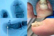
Diagnosis of nail exostosis is performed by an orthopedist or traumatologist. Sometimes the neoplasm can be identified already during the first medical consultation, but more often additional tests are prescribed to confirm the diagnosis - in particular, X-rays. In the X-ray image, the growth is somewhat smaller than it actually is, because the cartilage layer is not visualized on the image. In individual cases, a CT scan, magnetic resonance imaging, biopsy (if the growth is rapidly and intensively enlarging) may be required. To exclude a malignant process, the biomaterial is sent to the laboratory for subsequent cytologic analysis. [7]
It is mandatory to carry out differential diagnosis. Nail exostosis has similar symptomatology with other pathologies:
- Onychodystrophy (trophic disorder and thinning of the nail plate);
- Nail ingrowth;
- Mycosis (fungal lesions).
Many physicians can easily confuse exostosis with a dermatologic disease unless they refer the patient for radiography.
Some experts distinguish between real and false exostosis of the nail, although such a classification is not officially approved. False exostosis can be understood as a consequence of any trauma to the finger, mainly a fracture, in which there was improper fusion of bone fragments, which has the appearance of an outgrowth.
Who to contact?
Treatment of the subfoot exostosis
In the absence of symptoms and small size of the neoplasm, it is possible to establish dynamic monitoring of nail exostosis. Otherwise, the problem is solved exclusively by surgical removal. No conservative methods are not able to cause resorption of the formed exostosis and equalization of the nail plate. Taking analgesics, rubbing anti-inflammatory drugs is only a temporary way to improve the condition, but is not able to cure the disease.
The only radical method of getting rid of exostosis of the nail is surgical treatment. The operation is relatively simple, there is no need for prolonged recovery measures, a long stay in hospital. [8]
The intervention is minimally invasive and local anesthesia is used. On average, the procedure lasts about half an hour. On the same day, the patient can leave the clinic and go about his daily duties. Restrictions provide only a reduction in physical activity on the operated limb - on average, for 10-14 days. During this period, it is necessary to perform dressings, treatment of the affected finger with antiseptic solutions.
Closed shoes must not be worn during the rehabilitation period. Since the operated toe will be bandaged, sandals, flip-flops, soft slippers with open toes are allowed as footwear.
Removal of nail exostosis
Nail exostosis can only be completely cured with surgery. The surgeon excises the bone tissue and restores the normal configuration of the bone. The surgery involves a gentle method performed in stages:
- Defining the area of intervention, treating it with antiseptic solution to prevent wound infection.
- Performing anesthesia in the form of injection or application of an anesthetic drug.
- Vascular blockage (applying a tourniquet to prevent massive bleeding).
- Directly removing the exostosis.
- Suturing of the incision, re-treatment with antiseptic solution.
Surgical intervention is relatively uncomplicated, tissues recover quickly. There is no need to apply a plaster cast or use crutches. Upon completion of the operation, the surgeon bandages the operated finger: dressings should be performed regularly for several days, while treating the postoperative wound with recommended antiseptic solutions. During the entire rehabilitation period, it is necessary to visit the doctor, monitor the healing process, follow the rules of wound care. In general, recovery takes about 1.5-2 months.
Prevention
Preventive measures to prevent the development of nail exostosis can be as follows:
- Wearing comfortable shoes made of soft materials, with a wide toe and small heel for comfort and to minimize toe compression;
- Avoid wearing tight, tight shoes, shoes that are not the right size, have rough seams and elements that put pressure on any area of the foot and toes;
- Strengthening of the foot muscles, regular exercises (clenching and unclenching of the toes, circular and wiggling movements of the feet);
- Avoid overloading of the lower limbs, control of physical activity, avoid prolonged monotonous position of the feet;
- Weight control.
It is recommended to use special shoes for sports activities. You should not go jogging in ordinary shoes or sneakers that are not designed for training.
Forecast
The outcome of the disease can be considered favorable. In surgical treatment, the growth is removed, otherwise there is further progression of the pathology. In some situations, recurrences are possible. Malignization is likely for less than 1% of exostoses. Most often malignization is detected in patients with multiple bone and cartilage neoplasms. Malignant degeneration can be suspected with a sudden progression of growth of the neoplasm, a sharp increase in its diametric size.
After the nail exostosis is removed, the patient will have to undergo a rehabilitation course to restore the health of the nail plate. The course of postoperative treatment includes taking vitamin and mineral preparations, as well as medications that promote the regeneration of cartilage and bone tissue.
Literature
Saveliev, V. S. Clinical Surgery. In 3 vol. Vol. 1: national manual / Ed. By V. S. Saveliev. С. Savelyev, A. I. Kirienko. - Moscow: GEOTAR-Media, 2008.