Medical expert of the article

New publications
Teeth
Last reviewed: 07.07.2025

All iLive content is medically reviewed or fact checked to ensure as much factual accuracy as possible.
We have strict sourcing guidelines and only link to reputable media sites, academic research institutions and, whenever possible, medically peer reviewed studies. Note that the numbers in parentheses ([1], [2], etc.) are clickable links to these studies.
If you feel that any of our content is inaccurate, out-of-date, or otherwise questionable, please select it and press Ctrl + Enter.
Teeth (dentes) are important anatomical structures located in the dental alveoli of the jaws. Depending on the features of the structure, position and function, several groups of teeth are distinguished: incisors, canines, small molars, or premolars, and large molars.
Incisors are mainly used for grasping and biting off food, canines - for crushing it, molars - for grinding and grinding food. Despite the division of teeth into different groups, all teeth have a common structure. The tooth consists of a crown, neck and root.
The crown of the tooth (corona dentis), the most massive part protruding above the gum, has several surfaces. The lingual surface (facies lingualis) of the crown faces the tongue, the vestibular (facial) surface (facies vestibularis, seu facialis) faces the vestibule of the mouth, and the contact surface (facies contactus) faces the adjacent tooth. The chewing surfaces (facies masticatoria), or the occlusal surface (facies occlusiatis), of the analogous teeth of the upper and lower jaws face each other.
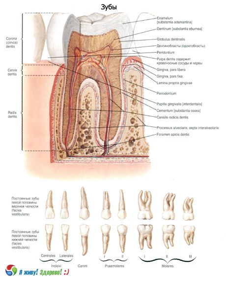
Inside the crown is the coronal cavity (cavitas coronalis), which contains the pulp and continues into the root canal of the tooth.
The root of the tooth (radix dentes) is located in the dental alveolus, with the walls of which it is connected by a special type of synarthrosis - hammering. Each tooth has from one (incisors, canines) to two or three (molars) roots. Inside each root there is a tooth canal (canalis radicis dentis), also filled with pulp. The root of the tooth ends with the apex (apex radicis dentis), which has an opening through which an artery and nerve enter the cavity of the tooth, and a vein exits.
Between the crown and the root is the neck of the tooth (cervix dentis), which is covered by the mucous membrane of the gum.
The dental pulp (pulpa dentis) is formed by loose fibrous connective tissue in which blood vessels and nerves branch out.
The main mass of the tooth is formed by dentin (dentinum). In the area of the crown, dentin is covered with enamel, in the area of the neck of the tooth and its root - with cement.
Enamel (enamelum) is a very durable substance. It is built of enamel prisms 3-5 µm thick, separated from each other by an interprismatic component. This component has a lower electron density compared to enamel. The free surface of the enamel is covered with a thin cuticle. Enamel mainly consists of inorganic salts (96-97%), among which calcium phosphate and calcium carbonate predominate. Enamel contains almost 4% calcium fluoride. Dentin contains about 28% organic substances (mainly collagen) and 72% inorganic substances. Among the inorganic compounds, calcium phosphate, magnesium phosphate and calcium fluoride predominate.
The structure of cement resembles bone tissue. It is formed by calcified plates, between which are multi-branched cementocytes located in lacunae. Collagen (Sharpey's) fibers penetrate the cement, which tightly fasten the root of the tooth to the periodontium. In the area of the neck of the tooth, the cement is eroded, devoid of cells (acellular cement). The composition of cement includes 29.6% organic substances and 70.4% inorganic compounds (mainly calcium phosphate and calcium carbonate).
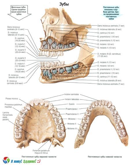
A distinction is made between milk teeth and permanent teeth.
Baby teeth (dentes decidui) appear in a child after birth, starting from 5-7 months of life, in the amount of 20. At the age of 5-7 years, baby teeth fall out and are replaced by permanent teeth (dentes permanentes), the number of which in an adult reaches 32. Baby teeth, compared to permanent teeth, have relatively wider and shorter crowns and short roots. A child has 2 incisors, 1 canine, 2 molars on each upper jaw bone and half of the lower jaw. Small molars are absent (0).
Timing of eruption of milk and permanent teeth
Tooth |
Jaw |
Timing of teething |
|
Dairy, months |
Constant, years |
||
Medial incisor |
Upper Lower |
7-8 5-7 |
7-8 6-7 |
Lateral incisor |
Upper Lower |
8-9 7-8 |
8-9 7-8 |
Fang |
Upper Lower |
18-20 16-18 |
11-12 9-10 |
First premolar |
Upper Lower |
- - |
10-11 10-12 |
Second premolar |
Upper Lower |
- - |
10-12 11-12 |
First molar |
Upper Lower |
14-15 12-13 |
6-7 6-7 |
Second molar |
Upper Lower |
21-24 20-22 |
12-13 11-13 |
Third molar |
Upper Lower |
- - |
17-21 12-26 |
In digital terms, the formula for milk teeth is as follows:
2012 |
2102 |
2012 |
2102 |
In this formula, the top row represents the upper teeth, the bottom row represents the lower teeth. The vertical line separates the teeth on the right side from the teeth on the left side. Each number represents the number of teeth of a certain shape.
Before a permanent tooth erupts, the corresponding baby tooth falls out. The eruption of permanent teeth begins at the age of 6-7 and continues until the age of 13-15. The first to erupt are the lower molars, then the medial incisors and the first upper molars, followed by the lateral incisors. Later, the first molars appear, followed by the canines, then the second premolars, and after them the second molars. The third molars, or wisdom teeth, are the last to erupt (at the age of 22-26). Each half of the upper jaw and each half of the lower jaw have 8 permanent teeth: 2 incisors, 1 canine, 2 premolars and 3 molars.
The dental formula of permanent teeth is as follows:
3212 |
2123 |
3212 |
2123 |
Incisors (dentes incisivi) have a flattened wide crown with a cutting surface. The crown of the upper incisors is wider than the lower ones. The root of the incisors is single, conical; in the lower incisors, the root is compressed from the sides. Depending on the location in relation to the median plane, lateral and medial incisors are distinguished.
Canines (dentes canini) have a conical, pointed crown. The root is single, long, compressed from the sides. The root of the lower canines is shorter than the upper ones. Sometimes the root of the lower canines is bifurcated.
Small molars (premolars - dentes premolares) are located behind the canine tooth. The crown of the premolars is round or oval from the chewing surface, has two chewing tubercles. The height of the crown is less than that of the canines. The root of the premolars is single, conical in shape, in the upper premolar it is sometimes bifurcated.
The large molars (dentes molares) are located behind the premolars. The crown of the large molars is usually cubic in shape, with 3-5 tubercles on the chewing surface. The large molars of the upper jaw have 3 roots, the lower - 2. The size of the molars decreases from front to back. The third molar (wisdom tooth - dens serotinus) is the smallest in size.
What do need to examine?