Medical expert of the article

New publications
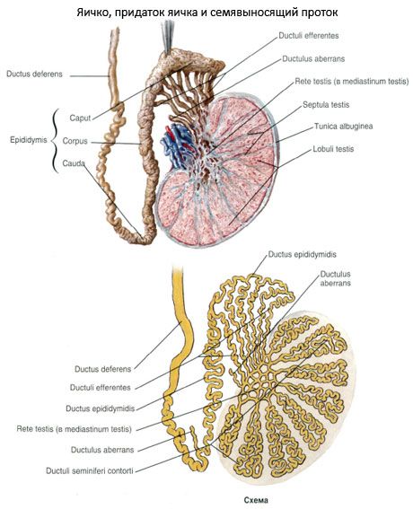
Seminiferous duct
Last reviewed: 07.07.2025

All iLive content is medically reviewed or fact checked to ensure as much factual accuracy as possible.
We have strict sourcing guidelines and only link to reputable media sites, academic research institutions and, whenever possible, medically peer reviewed studies. Note that the numbers in parentheses ([1], [2], etc.) are clickable links to these studies.
If you feel that any of our content is inaccurate, out-of-date, or otherwise questionable, please select it and press Ctrl + Enter.
The vas deferens is a paired organ, a direct continuation of the duct of the epididymis and ends at the point of confluence with the excretory duct of the seminal vesicle. The length of the vas deferens is about 50 cm, the diameter is about 3 mm, and the diameter of the lumen does not exceed 0.5 mm. The wall of the duct is significantly thick, so it does not collapse and is easily palpated as part of the spermatic cord.
Based on the topographic features of the vas deferens, it is divided into 4 parts. The initial, shortest section, located behind the testicle, medial to its appendage, is called the testicular part. The next part, rising vertically upward, passes as part of the spermatic cord, medial to its vessels, and reaches the superficial inguinal ring - this is the cord part. Then the vas deferens enters the inguinal canal, where its inguinal part is located. Having exited the inguinal canal through the deep inguinal ring, the vas deferens is directed along the lateral wall of the small pelvis downwards and backwards to merge with the excretory duct of the seminal vesicle. This section of the vas deferens is called the pelvic part. In the cavity of the small pelvis, the duct is located under the peritoneum (retroperitoneally). On its way it bends around the lateral side of the trunk of the inferior epigastric artery, crosses with the external iliac artery and vein, penetrates between the urinary bladder and the rectum, crosses the ureter, reaches the bottom of the urinary bladder and approaches the base of the prostate gland, next to a similar duct on the opposite side. This final section of the vas deferens is expanded, fusiform and forms the ampulla of the vas deferentis (ampulla ductus deferentis). The length of the ampulla is 3-4 cm, its largest transverse dimension reaches 1 cm. In the lower part, the ampulla gradually narrows and, entering the thickness of the prostate gland, connects with the excretory duct of the seminal vesicle.
The wall of the vas deferens consists of the mucous, muscular and adventitial membranes. The mucous membrane (tunica mucosa) forms 3-5 longitudinal folds. In the area of the ampulla of the vas deferens, the mucous membrane has bay-shaped protrusions - ampulla diverticula (diverticulum ampullae). Outside the mucous membrane is the muscular membrane (tunica muscularis). It consists of obliquely oriented middle circular, internal and external longitudinal layers of unstriated (smooth muscle) cells. The muscular membrane gives the wall of the vas deferens an almost cartilaginous density. In the ampulla of the vas deferens, the muscular layers are less distinct.
The external wall of the vas deferens is represented by the adventitial membrane (tunica adventitia), which passes without sharp boundaries into the connective tissue surrounding the duct.
What tests are needed?