Medical expert of the article

New publications
Prostate gland (prostate)
Last reviewed: 04.07.2025

All iLive content is medically reviewed or fact checked to ensure as much factual accuracy as possible.
We have strict sourcing guidelines and only link to reputable media sites, academic research institutions and, whenever possible, medically peer reviewed studies. Note that the numbers in parentheses ([1], [2], etc.) are clickable links to these studies.
If you feel that any of our content is inaccurate, out-of-date, or otherwise questionable, please select it and press Ctrl + Enter.
The prostate gland (prostata, s.glandula prostatica, prostate) is an unpaired muscular-glandular organ. The gland secretes a secretion that is part of sperm. The secretion liquefies sperm, promotes sperm motility.
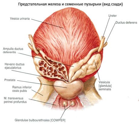
The prostate gland is located in the anteroinferior part of the small pelvis under the urinary bladder, on the urogenital diaphragm. The initial section of the urethra, the right and left ejaculatory ducts pass through the prostate gland.
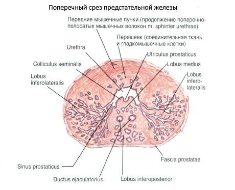
The shape of the prostate gland resembles a chestnut, slightly flattened in the anteroposterior direction. The prostate gland has an upward-facing base (basis prostatae), which is adjacent to the bottom of the urinary bladder, seminal vesicles and ampullae of the vas deferens. The anterior, posterior, lower-lateral surfaces and the apex of the gland are also distinguished.
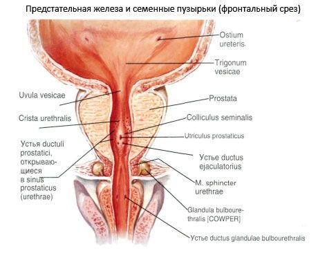
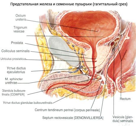
The anterior surface (facies anterior) faces the pubic symphysis and is separated from it by loose tissue with the venous plexus located in it. The lateral and median puboprostatic ligaments (ligg.puboprostaticae) and the puboprostatic muscle (m.puboprostaticus) go from the prostate gland to the pubic symphysis. The posterior surface (facies posterior) is directed toward the ampulla of the rectum and is separated from it by a connective tissue plate - the rectovesical septum (septum rectovesicale). The proximity to the rectum allows the prostate gland to be palpated in a living person through the anterior wall of the rectum. The inferolateral surface (facies inferolateralis) is rounded and faces the muscle that lifts the anus. The top of the prostate gland (apex prostatae) faces downwards and is adjacent to the urogenital diaphragm. The urethra enters the base of the prostate gland, with most of the gland remaining behind the canal, and exits the gland in the area of its top. The transverse size of the prostate gland reaches 4 cm, the longitudinal (upper-lower) is 3 cm, the anteroposterior (thickness) is about 2 cm. The mass of the gland is 20-25 g.
The substance of the prostate gland has a dense consistency and a grayish-red color. The prostate gland has two lobes: the right lobe (lobus dexter) and the left lobe (lobus sinister). The border between them is visible on the anterior surface of the gland as a shallow groove. The part of the gland protruding on the posterior surface of the base and limited by the urethra in front and the ejaculatory ducts behind is called the isthmus of the prostate gland (isthmus prostatae), or the middle lobe (lobus medius). This lobe often hypertrophies in old age and makes urination difficult.
 [
[ Structure of the prostate gland
The prostate gland is covered externally by a capsule (capsula prostatica), from which bundles of connective tissue fibers - the septa of the prostate gland - branch off into the gland. The parenchyma (parenchyma) consists of glandular tissue, as well as smooth muscle tissue, which makes up the muscular substance (substantia muscularis). The glandular tissue is grouped into separate complexes in the form of glands (lobules) of an alveolar-tubular structure. The number of glandular lobules reaches 30-40. They are located mainly in the posterior and lateral parts of the prostate gland. There are few lobules in the anterior part of the prostate gland. Small mucous glands opening into the urethra are located directly around the urethra. Smooth muscle tissue prevails here, which is concentrated around the lumen of the male urethra. This muscular tissue of the prostate gland unites with the muscular bundles of the bottom of the urinary bladder and participates in the formation of the internal (involuntary) sphincter of the male urethra. The glandular passages of the glands, merging in pairs, pass into the excretory prostatic ducts (ductuli prostatici), which open into the male urethra in the area of the seminal hillock with pinpoint openings. Contraction of the muscular bundles facilitates the removal of the secretion of the prostatic and mucous glands into the urethra.
Vessels and nerves of the prostate gland
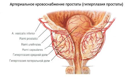
The blood supply to the prostate gland is provided by numerous small arterial branches originating from the inferior vesical and middle rectal arteries (from the system of internal iliac arteries). Venous blood from the prostate gland flows into the venous plexus of the prostate gland, from there into the inferior vesical veins, which flow into the right and left internal iliac veins. The lymphatic vessels of the prostate gland flow into the internal iliac lymph nodes.
The nerves of the prostate gland originate from the prostatic plexus, which receives sympathetic (from the sympathetic trunks) and parasympathetic (from the pelvic visceral nerves) fibers from the inferior hypogastric plexus.