Rectum
Last reviewed: 31.05.2018

All iLive content is medically reviewed or fact checked to ensure as much factual accuracy as possible.
We have strict sourcing guidelines and only link to reputable media sites, academic research institutions and, whenever possible, medically peer reviewed studies. Note that the numbers in parentheses ([1], [2], etc.) are clickable links to these studies.
If you feel that any of our content is inaccurate, out-of-date, or otherwise questionable, please select it and press Ctrl + Enter.
The rectum is the final section of the large intestine. Its length averages 15 cm, and its diameter ranges from 2.5 to 7.5 cm. The rectum is divided into two sections: the ampulla and the anal canal. The ampulla of the rectum (ampula recti) is located in the pelvic cavity, and the anal canal (canalis analis) is located in the perineum. The sacrum and coccyx are located behind the ampulla. In front of the rectum in men are the prostate gland, bladder, seminal vesicles, and ampulla of the right and left vas deferens, and in women, the uterus and vagina. The anal canal ends in the anus.
The rectum forms bends in the sagittal plane. The upper - sacral bend (flexura sacralis), facing backwards with its convexity, corresponds to the concavity of the sacrum. The lower - perineal bend (flexura perineals), directed forwards, is located in the thickness of the perineum (in front of the coccyx). The bends of the rectum in the frontal plane are inconstant. The upper part of the intestine is covered by the peritoneum on all sides, the middle - on three sides, the lower part does not have a serous cover.
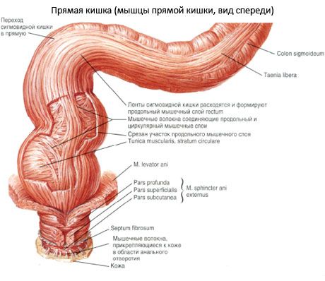
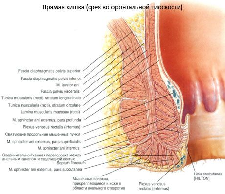
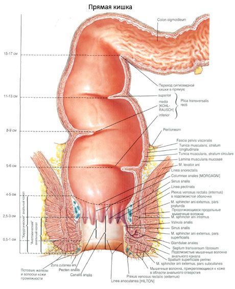
In the anal canal area, the thickening of the muscular membrane of the intestine forms the internal (involuntary) sphincter of the anus (m.sphincter ani internus). Directly under the skin is the external (voluntary) sphincter (m.sphincter ani extemus), formed by striated muscle fibers. It is part of the perineal muscles. Both sphincters close the anus and open during defecation.
The peritoneum on the sides of the rectum forms the sacrouterine folds. Between the latter and the lateral walls of the pelvis there are pelvic-rectal fossae. In the subperitoneal tissue of these fossae pass the ureters and branches of the hypogastric vessels, and in the fossae themselves lie the tubes and ovaries.
In front, the rectum in the subperitoneal space of the pelvic cavity is adjacent to the vagina. The peritoneal-perineal aponeurosis in women is a loose plate that allows the rectum to be easily separated from the vagina.
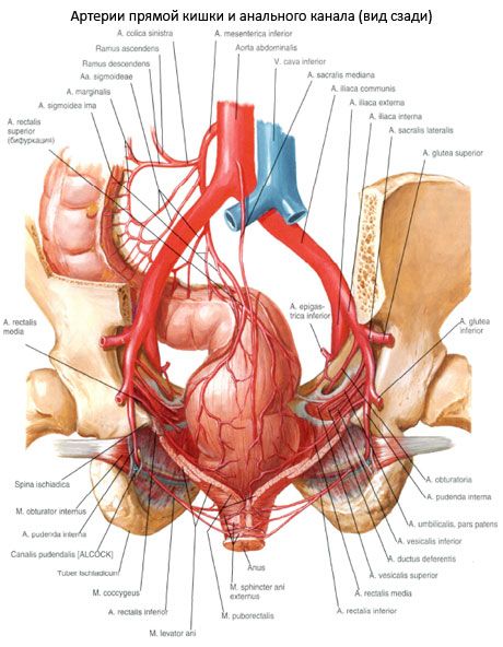
The rectum is supplied with blood by one unpaired artery - the superior rectal, which is the terminal branch of the inferior mesenteric artery, and two paired arteries - the middle rectal (a branch of the internal iliac artery) and the inferior rectal (a branch of the internal pudendal artery). The arterial trunks have a longitudinal direction in relation to the intestinal wall.
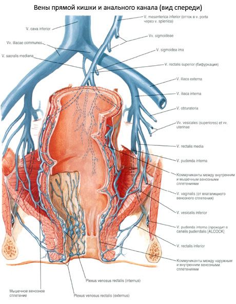
Venous outflow from the rectum goes into two venous systems - the inferior vena cava and the portal vein. In this case, three venous plexuses are formed: subcutaneous, submucous and subfascial. From the upper two-thirds of the rectum, venous blood flows through the upper rectal veins into the inferior mesenteric vein from the portal vein system, and from the lower third - into the inferior vena cava system.
Lymphatic drainage from the rectum occurs in four main directions:
- from the lower rectum to the inguinal lymph nodes;
- from the upper sections to the sacral lymph nodes;
- from the anterior sections to the upper rectal lymph nodes;
- from the middle sections to the lower iliac collectors.
Innervation of the rectum is carried out by sympathetic and parasympathetic (motor and sensory) fibers. Sympathetic fibers originate from the inferior mesenteric, aortic plexuses and reach the rectum either along the branches of the superior rectal artery or as part of the hypogastric nerves. The perineal part of the rectum is innervated by the genital nerve, which contains motor and sensory fibers.

 [
[