Medical expert of the article

New publications
Abdominal exam
Last reviewed: 04.07.2025

All iLive content is medically reviewed or fact checked to ensure as much factual accuracy as possible.
We have strict sourcing guidelines and only link to reputable media sites, academic research institutions and, whenever possible, medically peer reviewed studies. Note that the numbers in parentheses ([1], [2], etc.) are clickable links to these studies.
If you feel that any of our content is inaccurate, out-of-date, or otherwise questionable, please select it and press Ctrl + Enter.
For examination and further investigation of the abdomen, it must be sufficiently exposed. It is necessary that the groin areas be fully examined. The patient must lie in a comfortable position. The room must be warm.
Examination of the abdomen
On the skin of the abdomen, so-called striae (whitish stripes when stretched by edematous fluid or reddish-brown with hypercorticism) and superficial veins, the increased development of which is associated with liver pathology (collaterals in portal hypertension ) may be visible.
The abdomen is involved in the act of breathing, the absence of respiratory movements is characteristic of acute peritonitis. In the epigastric region, pulsation of the abdominal aorta may be visible, less often it is caused by a hypertrophied right ventricle of the heart.
During the examination, the shape and symmetry of both halves of the abdomen are assessed. The abdomen may be enlarged due to obesity, massive gas formation in the intestine, ascites, pregnancy, a large ovarian cyst, and sometimes an enlarged gallbladder. Swelling and deformation of the abdomen, visible during external examination, are possible due to the presence of tumors of various localizations, enlarged liver, spleen, and kidneys. Normal peristalsis of the small intestine is sometimes visible through the thin abdominal wall. Hernias of various localizations can cause local bulging of the abdominal wall. This applies to umbilical hernia, hernia of the white line of the abdomen, as well as femoral and inguinal hernias.
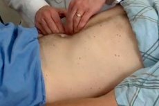
Palpation of the abdomen
It is important that the doctor's hands are warm. To relax the muscles of the anterior abdominal wall, the patient should be in a comfortable position with a low-lying head and arms extended along the body.
Superficial palpation begins with both hands, comparing symmetrical areas of the abdomen (pain, muscle tension, etc.). Then, placing the entire palm on the abdomen, the doctor begins to palpate the abdomen with the fingertips of the right hand, starting with the areas most distant from the site of pain. When moving the hand over the surface of the abdomen, the tension of the abdominal wall, hernial openings, divergence of the muscles of the abdominal wall, and pain when palpating certain parts of the abdomen are determined more accurately. Palpation as one of the main methods of physical examination of the abdominal organs has been widely used since the end of the last century, when in 1887 the Russian clinician V. P. Obraztsov first described in detail the results of targeted palpation of the abdomen. "Palping the patient's abdomen in a horizontal position," writes V. P. Obraztsov, "I felt three fingers below the navel, along the midline, the intestine in the form of a rather thick, mobile up and down, non-rumbling cylinder, which could be quite clearly traced to the right and left rising to the hypochondrium and disappearing behind them. With the same clarity and distinctness... I also felt two other cylinders descending on the sides, in a downward direction, of which one, the left, passed into the sigmoid colon, and the other, the right, into the cecum."
V. P. Obraztsov gives important methodological advice (which is the basis of the method he proposed for examining abdominal organs): place your hands with slightly bent fingers on either side of the navel and begin to move them up and down along with the abdominal walls.
This method of direct physical examination is called "methodical deep sliding palpation" because it combines the results of sensation obtained by the doctor simultaneously from static (contact with the skin of the abdomen and the wall of the organ) and dynamic (penetration of the doctor's hand or fingers deep, pressing on the underlying organ and sliding) palpation. The immersion of the fingers should be carried out gradually, during each exhalation of the patient, which allows for a maximum reduction in the reflex tension of the abdominal muscles and pressing the organ being examined to the back wall of the abdominal cavity: further palpation occurs with finger movements carried out in a direction perpendicular to the axis of the organ being palpated. When carrying out these movements, it is necessary to move the fingers together with the skin of the abdomen and underlying tissues. It is better to start palpation from the most accessible section - the sigmoid colon, then move on to the cecum, ileum, ascending, descending, transverse colon, palpate the liver, spleen.
The sigmoid colon can be palpated in all healthy people, except for those with large fat deposits. The sigmoid colon is normally palpated as a dense, smooth cylinder, about the thickness of a thumb. It is usually painless, and there is no rumbling.
The cecum is palpated in the right iliac region as a painless cylinder, two fingers thick. An attempt to palpate other parts of the large intestine is also possible: the ascending, descending and transverse colon. More often, palpation of them remains ineffective. With denser contents, these parts of the intestine can be palpated as dense strands.
The greater curvature of the stomach can be determined as a ridge. It is found by palpating the epigastric region at different levels. The pylorus is palpated to the right of the spine as a cord of varying density. With pathological changes, the pylorus becomes denser and more painful. Most often, the sections of the stomach are not palpated. However, many patients may experience not only pain in certain areas of the epigastric region during palpation, but also tension in the muscles of the abdominal wall (muscle protection), which is typical for gastric ulcer. Palpation of the stomach sometimes allows you to detect a tumor.
Percussion of the abdomen
The main purpose of abdominal percussion is to determine to what extent the abdominal enlargement is associated with the presence of gas, fluid, or a dense formation. A tympanic sound is characteristic of bloating associated with gas formation. Dullness of the percussion sound is usually noted with ascites. In these cases, the abdomen is often enlarged in volume, and the percussion sound becomes dull in the lateral parts of the abdomen. When the patient is turned on his side, tympanitis begins to be determined on the opposite side, which is associated with the movement of fluid into the lower parts of the abdominal cavity.
Palpation of the rectum is performed using the index finger inserted into the rectum through the anus ( digital examination of the intestine ). This allows one to establish the presence of hemorrhoids, tumors in the rectum, and also to palpate the prostate gland, uterus, ovaries, and infiltrates in the abdominal cavity bordering the rectum.
Auscultation of the abdomen
Intestinal peristalsis produces sounds that can be heard when a stethoscope is applied to the abdomen. Most often, these sounds are heard every 5-10 seconds, but these intervals may vary. Intestinal peristalsis disappears with intestinal obstruction due to obstruction of the intestinal lumen. The appearance of arterial noises when listening to the aorta and renal arteries at the point of their projection is associated with their narrowing. Occasionally, friction noises are heard, reminiscent of pleural friction noise in dry pleurisy, caused by the presence of perisplenitis or perihepatitis.
Additional research methods
Stool examination. Includes examination for occult blood, microscopy and bacteriological examination.
The occult blood test is important for the diagnosis of gastrointestinal and hematological diseases. A positive result may be the only initial sign of a colon tumor, since bleeding is episodic and it is necessary to obtain the results of at least three studies over several days. In the presence of hemorrhoids, it is advisable to obtain the material for the study using a proctoscope.
The most suitable test is the guaiac resin test. When oxidized, guaiac resin turns blue due to the activity of hemoglobin, similar to peroxidase.
During microscopic examination, a particle of feces is mixed on glass with a drop of isotonic sodium chloride solution. Microscopy can reveal erythrocytes and macrophages, which are found in large quantities in ulcerative lesions of the colon. Cysts and parasite eggs, undigested meat fibers can also be detected.
During a bacteriological study, quite a variety of microorganisms are always found in significant quantities in feces. Changes in the ratio of their proportions are noted in the so-called dysbacteriosis. In this case, a special quantitative study of feces is carried out for the presence of bacteria.
Endoscopic examination. The advent of gastrofibroscopes has significantly expanded the use of the endoscopic method for diagnosing diseases of various parts of the digestive tract. In this case, the rectum and the lower part of the sigmoid colon are examined using a solid endoscope. For examination of the colon, the patient must be sufficiently prepared (it is necessary to clean the intestines with enemas). During the examination, in addition to examination, a biopsy of the altered tissues is performed for microscopic examination. Currently, relatively small pathological formations, such as polyps, are removed through endoscopes.
X-ray examination. First, a general image of the abdominal cavity is taken, which may show the kidneys, less often the spleen, sometimes stones in the kidneys and bile ducts, less often phleboliths in the small pelvis. Of particular importance is an image of the abdomen in a standing and lying position for assessing the so-called acute abdomen. This allows us to detect the level of fluid and the distribution of gas along the gastrointestinal tract.
Contrast radiography allows us to clarify the condition of the digestive tract. When swallowing a barium suspension, we can detect a narrowing or widening of the esophagus in one section or another. Filling defects caused by a tumor or ulceration of the mucous membrane can be detected in the stomach. The duodenum and other sections of the small intestine are examined.
The colon is examined by introducing a barium suspension using an enema. The patient's preparation consists of a complete cleansing of the colon using laxatives and enemas. Sometimes this causes certain difficulties and unpleasant sensations for the patient and serves as a relative contraindication to this procedure.
When performing an X-ray examination of the gastrointestinal tract, it is important to prepare the patient, which consists of following a diet for 2-3 days before the procedure. In this case, products that cause strong gas formation (fresh milk, peas, cabbage and other vegetables) are excluded.
Endoscopy and contrast radiography of the digestive tract are considered complementary studies. When an accurate diagnosis is established with the help of one of them, the other is not required. Both methods are used when the diagnosis is in doubt and in any case when there is a suspicion of a tumor that may require surgical intervention.
Ultrasound examination. This method is used to obtain a two-dimensional image of the abdominal cavity, in particular dense organs such as the liver, spleen, kidneys, and abdominal lymph nodes.
Computer tomography. The method is used to assess the size of dense formations in the abdominal cavity, in particular the pancreas.
 [ 9 ]
[ 9 ]