Medical expert of the article

New publications
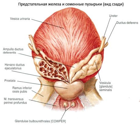
Seminal vesicle
Last reviewed: 07.07.2025

All iLive content is medically reviewed or fact checked to ensure as much factual accuracy as possible.
We have strict sourcing guidelines and only link to reputable media sites, academic research institutions and, whenever possible, medically peer reviewed studies. Note that the numbers in parentheses ([1], [2], etc.) are clickable links to these studies.
If you feel that any of our content is inaccurate, out-of-date, or otherwise questionable, please select it and press Ctrl + Enter.
The seminal vesicle (vesicula, s.glandula seminalis) is a paired organ located in the pelvic cavity lateral to the ampulla of the vas deferens, above the prostate gland, behind and to the side of the bottom of the urinary bladder. The seminal vesicle is a secretory organ. Its glandular epithelium secretes a secretion containing substances necessary for the nutrition and activation of spermatozoa.
The peritoneum covers only its upper sections. The surface of the seminal vesicle is tuberous. The seminal vesicle has an anterior surface facing the urinary bladder and a posterior surface adjacent to the rectum. The length of the seminal vesicle is about 5 cm, the width is 2 cm, and the thickness is 1 cm. When cut, it looks like communicating vesicles. If the outer membrane of the seminal vesicle is partially removed and straightened, it takes the form of a tube 10-12 cm long and 0.6-0.7 cm thick.
On the outside, the seminal vesicle has an adventitial membrane (tunica adventitia). Inside, there is a well-developed muscular membrane (tunica muscularis), represented by two layers of smooth myocytes. The bundles of the inner layer have a circular orientation, those of the outer layer - longitudinal.
The mucous membrane (tunica mucosa) forms longitudinal folds that significantly increase the surface area of the secretory epithelium of the seminal vesicle. The epithelial cover is formed by tall, cylindrical secretory cells on a thin basement membrane. Each seminal vesicle has an upper, widened end - the base, a middle part - the body and a lower, tapering end that passes into the excretory duct (ductus excretorius). The excretory duct of the seminal vesicle connects with the final section of the vas deferens and forms the ejaculatory duct (ductus ejaculatorius), which pierces the prostate gland and opens into the prostatic part of the male urethra, on the side of the seminal hillock. The length of the ejaculatory duct is about 2 cm, the width of the lumen is from 1 mm at the initial part to 0.3 mm at the point of entry into the urethra.
The mucous membrane of the ejaculatory duct forms longitudinal folds and is covered with prismatic epithelium containing pigment granules. At the point where the duct passes through the prostate gland, its muscular membrane passes into the muscles of this gland.
Vessels and nerves of the seminal vesicle and vas deferens. The seminal vesicle is supplied with blood from the descending branch of the artery of the vas deferens (a branch of the umbilical artery). The ascending branch of the artery of the vas deferens brings blood to the walls of the vas deferens. The ampulla of the vas deferens receives blood through branches of the middle rectal artery and the inferior vesical artery (from the internal iliac artery).
Venous blood from the seminal vesicles flows through the veins into the venous plexus of the urinary bladder and then into the internal iliac vein. Lymph from the seminal vesicles and the vas deferens flows into the internal iliac lymph nodes. The seminal vesicles and the vas deferens receive sympathetic and parasympathetic innervation from the plexus of the vas deferens (from the inferior hypogastric plexus).
 [
[