Medical expert of the article

New publications
Testicular appendage
Last reviewed: 06.07.2025

All iLive content is medically reviewed or fact checked to ensure as much factual accuracy as possible.
We have strict sourcing guidelines and only link to reputable media sites, academic research institutions and, whenever possible, medically peer reviewed studies. Note that the numbers in parentheses ([1], [2], etc.) are clickable links to these studies.
If you feel that any of our content is inaccurate, out-of-date, or otherwise questionable, please select it and press Ctrl + Enter.
The epididymis is located along the posterior edge of the testicle. There is a rounded, widened upper part - the head of the epididymis (caput epididymidis), which passes into the middle part - the body of the epididymis (corpus epididymidis). The body of the epididymis continues into a tapering lower part - the tail of the epididymis (cauda epididymidis). On the head of the epididymis there is an appendage of the epididymis (appendix epididymidis) in the form of a vesicle on a stalk, which is a rudimentary outgrowth of the mesonephric duct. In the area of the head and tail of the epididymis there may be blind-ending tubes - deflecting ducts (ductuli aberrantes) - the remains of the canals of the mesonephros (Wolffian body).
Behind the head of the appendage, in the connective tissue, there is a flat whitish formation, well expressed in children - the appendage of the testicle (paradydymis), also a rudiment of the mesonephros.
The serous membrane covering the testicle also extends onto the epididymis, and on the lateral side it enters the depression between the testicle and the epididymis, lining the sinus of the epididymis (sinus epididymidis). The efferent ducts of the testicle, which have a tortuous course, form conical lobules (cones) of the epididymis (lobuli epididymidis) in the epididymis, separated by thin connective tissue septa. There are 12-15 lobules in the epididymis. Each duct of the lobule flows into the duct of the epididymis (ductus epididymidis), which forms numerous bends along the entire length of the epididymis. The duct of the epididymis in a straightened form is 6-8 m long. In the caudal part of the epididymis, its duct passes into the vas deferens.
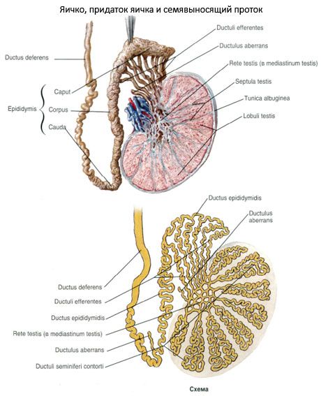
The mucous membrane of the duct of the epididymis is lined with pseudo-stratified (multi-row) columnar epithelium. The columnar epithelial cells have cytoplasmic outgrowths (stereocylls) on the apical surface. Between the basal part of the columnar epithelial cells are intercalated cells. The epithelium of the duct of the epididymis is located on the basement membrane. It participates in the formation of fluid that facilitates the passage of spermatozoa through the vas deferens. The epithelial cells also produce glycocalyx, which covers the spermatozoa with a thin layer. At the same time, the epididymis is a reservoir where spermatozoa accumulate, where they mature biochemically. When leaving the epididymis, the spermatozoa, however, are not fully mature and ready for fertilization.
Male reproductive cells (spermatozoa) are produced only in the convoluted seminiferous tubules of the testicle. All other tubules and ducts of the testicle and epididymis are the vas deferens. Spermatozoa are part of semen, the liquid part of which is represented by the secretion of the seminal vesicles and prostate gland.
 [
[ What tests are needed?