Medical expert of the article

New publications
The spermatic cord
Last reviewed: 07.07.2025

All iLive content is medically reviewed or fact checked to ensure as much factual accuracy as possible.
We have strict sourcing guidelines and only link to reputable media sites, academic research institutions and, whenever possible, medically peer reviewed studies. Note that the numbers in parentheses ([1], [2], etc.) are clickable links to these studies.
If you feel that any of our content is inaccurate, out-of-date, or otherwise questionable, please select it and press Ctrl + Enter.
The spermatic cord (funiculus spermaticus) is formed during the descent of the testicle. It is a round cord 15-20 cm long, extending from the deep inguinal ring to the upper end of the testicle. From the inguinal canal under the skin of the pubic region, the spermatic cord exits through the superficial inguinal ring. The spermatic cord includes the vas deferens, testicular artery, artery of the vas deferens, pampiniform (venous) plexus, lymphatic vessels of the testicle and its appendage, nerves, as well as traces (remnants) of the vaginal process in the form of a thin fibrous cord.
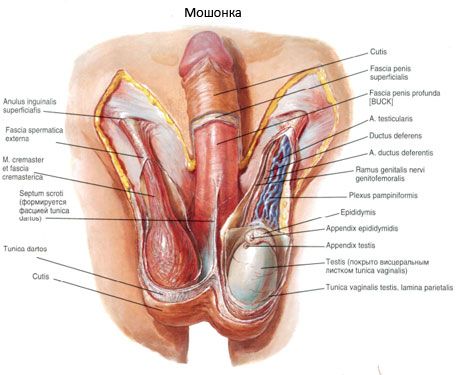
The vas deferens, which is the main component of the spermatic cord, as well as the vessels and nerves, are surrounded by membranes (tunicae funiculi spermatici), which continue into the membranes of the testicle. The innermost membrane, directly enveloping the duct, vessels and nerves, is the internal spermatic fascia (fascia spermatica interna). Outside of it are the muscle that lifts the testicle (m.cremaster) and the fascia of this muscle (fascia cremasterica). The outermost membrane of the spermatic cord is the external spermatic fascia (fascia spermatica externa), which envelops the entire spermatic cord from the outside.
 [ 1 ]
[ 1 ]
What do need to examine?